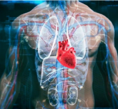
January 29, 2016 — Diseased hearts may be thrown out of rhythm by structural differences, now visible for the first time, in protein groups that connect heart muscle cells, according to the authors of a study published in the journal Nature Communications online.
By combining powerful imaging techniques and mathematical models, a research team led by NYU Langone Medical Center was able to reconstruct for the first time detailed, 3-D images in mice of intercalated discs, protein structures that connect heart muscle cells in working groups. These discs pass on both the electrical signals and the pumping force needed for the heart to beat normally.
The study found that the proteins that do these two jobs occur together in clusters. Conversely, instances where these proteins are farther apart than normal, even by billionths of a meter, may represent a newfound spatial signature of electrical malfunction.
“Our new images could someday help physicians and genetic counselors more accurately identify people at risk before they develop potentially life-threatening arrhythmias, electrical disorders that throw off the heart’s rhythm,” said senior study investigator Mario Delmar, M.D., Ph.D., the Patricia and Robert Martinsen Professor of Cardiology in the Department of Medicine’s Leon H. Charney Division of Cardiology at NYU Langone. His long-term goal is to develop a simple blood test that could detect dangerous disc protein structures as part of mass screening.
Presently, many arrhythmias go undiagnosed. Such conditions cause no symptoms in some, while others may be prone to fainting. In rare cases, arrhythmias can be fatal when they cause the heart to stop beating. And while electrical defects can be detected by an electrocardiogram (EKG), most people do not undergo such tests unless there is a known pattern of risk in their family.
“Assessing risk for arrhythmia based on real-time, structural analysis instead of guesswork based on heredity would represent a major advance,” said Delmar.
“We used innovative combinations of imaging techniques to visualize previously unseen aspects of the discs,” said Eli Rothenberg, Ph.D., an assistant professor at NYU Langone and corresponding author of the paper along with Delmar. “Focused ion beam-scanning electron microscopy, for instance, helped us to create 3-D images at nanometer resolution, which is down to one-billionth of a meter.”
Past studies have shown that intercalated discs consist partly of cadherins, proteins that anchor one cell to the next so they can pass on combined pumping force (adhesion). Also within the discs are protein channels that, upon receiving the right signal, let charged sodium particles flow through them. This flow serves as a switch that triggers heart muscle cell contraction (excitability).
Going into the current study, it was thought that adhesion and excitability were carried out by separate groups of disc molecules. But, mounting data argue that they work together. Specifically, Delmar and his colleagues revealed the existence in the discs of distinct clusters of both the adhesion molecule N-Cadherin and a protein, Nav 1.5, which is linked to sodium ion channels.
The authors speculate the clusters identified by the study are the sites of “crosstalk” between the contractile and electrical machinery in heart muscle. Their experiments revealed, for instance, that the ability of the discs to pass on electrical signals may also equip them to more strongly adhere to each other. This would extend the study implications to heart failure, say the authors, if structural problems in discs can be tied to lower adherence, less ability to pass on force, and a weaker heartbeat.
Most promising, the researchers say, is that their discovery of the proximity of adhesion and excitability complexes may represent a new way to measure disease risk through imaging, especially in cases where disease-causing genetic changes (mutations) can be linked to differences in protein spacing. Study results showed that about 60 percent of the N-cadherin in the intercalated discs of study mice was clustered within 100 nanometers of Nav 1.5.
Rothenberg said images of the proteins — tagged with a fluorescent dye to make them light up — showed “a highly organized distribution.” Nano-scale images revealed groups of 50 to 60 sodium channels arranged along the borders between cells and their intercalated discs where N-cadherin and Nav. 1.5 were concentrated. Similar clustering had been seen before in nerve cells, but never in heart muscle.
“The closeness of the proteins is probably essential to coordinating the electrical properties of the heartbeat,” said Rothenberg.
As the next step, the team plans experiments with human heart cells to see if intercalated disc malformations can be linked to arrhythmias.
Funding for the study, which took four years to complete, was provided by National Heart, Lung and Blood Institute (NHLBI) grant RO1 HL106632 and by National Institute of General Medical Sciences (NIGMS) grants RO1 GM57691, RO1 GM110385, RO1 GM108119; and R21 CA187612. Both NHLBI and NIGMS are part of the National Institutes of Health.
Other researchers at NYU Langone involved in this study were Alejandra Leo-Macias, Ph.D.; Esperanza Agullo-Pascual, Ph.D.; Sara Keegan, Ph.D.; Xianming Lin, Ph.D.; Fengxia Liang, Ph.D.; and David Fenyo, Ph.D., who provided the mathematical modeling needed to analyze the data. Additional research support was provided by Jose Sanchez-Alonso, Ph.D.; Yuri Korchev, Ph.D.; and Julia Gorelik, Ph.D., at the Imperial College in London, United Kingdom.
For more information: www.nature.com/ncomms

 November 12, 2025