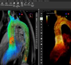
July 3, 2017 — When diagnosing strokes and heart diseases or looking at tumors, perfusion magnetic resonance imaging (MRI) offers a gentler way to capture the blood flow circulation in the organs. However, the method is far from being implemented to its full potential at many clinics. The Fraunhofer Institute for Medical Image Computing MEVIS in Bremen, Germany organized a workshop entitled “Measurement of Perfusion and Capillary Exchange” from June 21-23 to promote adoption of the method. The event provided information about its applications and the current state of research.
MRI allows a gentler way of taking 3-D images of the inside the body. One specific variant is perfusion MRI. It visualizes the perfusion of organs and tumors. “Perfusion measurements deliver important information about an organ’s condition,” said Fraunhofer MEVIS researcher and workshop initiator Matthias Günther. “It enables clinicians to diagnose at an early stage how well an organ is functioning.”
Physicians can also measure the perfusion with other methods, such as ultrasound, computed tomography (CT) and positron emission tomography (PET), but perfusion MRI offers several advantages. Unlike ultrasound, it visualizes the blood flow through even very fine blood vessels. Other than CT and PET, this method is not based on X-rays or radioactive substances that can damage the tissue.
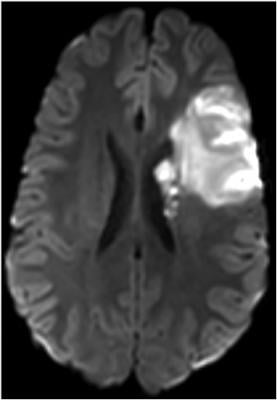
Today, perfusion MRI is primarily applied when diagnosing strokes and tumors. It measures which brain areas receive insufficient blood supply after a stroke occurs. It can also assess the viability and functionality of the liver and kidneys. Furthermore, the method can visualize which regions of a tumor are supplied with the most blood. In these areas, the ulcer grows the fastest, but is also more receptive to therapeutic treatment – helpful information for radiation therapists.
To perform perfusion MRI, physicians usually administer a contrast agent, which flows through the vessels with the blood and can be seen on the MR images due to its contrast. Among other things, the method can prove if a blood vessel in the brain has become porous due to a disease. In such cases, the contrast agent molecules can slip through the pores. The workshop was designed to bring the participants – primarily physicists, engineers, computer scientists and physicians – up to date and give them a foundation to further develop and apply the techniques in the clinical workflow.
A new alternative that does not require contrast agents has recently emerged, called arterial spin labeling. An MR scanner magnetically tags the blood flowing through an organ. The scanner ‘flips’ the original orientation of the atoms in the magnetic field. Then, it follows the magnetically tagged blood on its way through the brain vessels. “The method without contrast agents is non-invasive and less stressful for patients,” said Günther. “For patients who are frequently examined, some contrast agents run the risk of accumulating in the body.”
In recent years, the researchers have made advances in developing the method without contrast agents. They were able to increase its efficiency and reduce the examination time from 15 to between 3 and 5 minutes. “As scientists, we believe that the method is now ready for clinical use,” said Günther. Still, the method is currently only sporadically used in the clinical workflow.
“Despite its advantages, many clinics haven’t yet ventured into using the method,” explained Günther. “Our workshop should help ease concerns and contribute to making the method easier for physicians to apply.” Admittedly, operation and use are more challenging than standard MRI, and handling the new technique requires a certain degree of training. “With our workshop, we want to convince prospective users that learning the new method is worth the time invested.”
For more information: www.esmrmb.org

 January 21, 2026