Technology | PET-CT | October 05, 2016

GE Healthcare Announces FDA Clearance of Discovery MI PET/CT System

New digital PET/CT system enables clinicians to detect smaller lesions

GE Healthcare, Discovery MI PET/CT system, FDA clearance, RSNA 2016

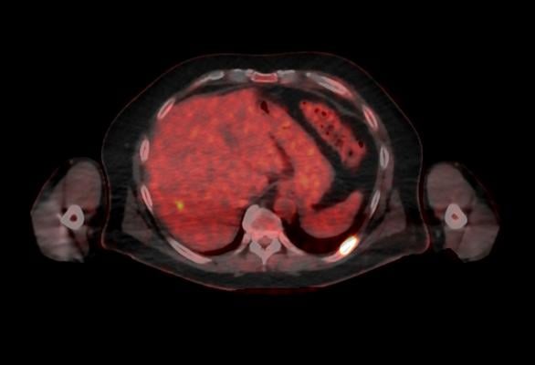
October 5, 2016 — GE Healthcare recently announced U.S. Food and Drug Administration (FDA) 510(k) clearance of its Discovery MI digital positron emission tomography (PET)/computed tomography (CT) system and shared a series of first clinical images. Built with technology allowing significantly better small lesion detectability, Discovery MI can help clinicians in their efforts to diagnose and stage disease earlier.

The first unit has been installed at the Stanford University School of Medicine, Stanford, Calif.  

Discovery MI was created to help clinicians achieve their primary goal of diagnosing and staging disease earlier and to better guide their treatment strategies. It also enables more compelling research with more novel, faster decaying tracers. A key component in delivering these desired outcomes is Discovery MI’s LightBurst Digital Detector. This detector delivers up to two times improvement in volumetric resolution, enabling small lesion detectability, and has the highest NEMA sensitivity of any time-of-flight (TOF)/PET system in the industry, according to GE.

Discovery MI may also enhance clinicians’ diagnostic service offerings in oncology and expand PET’s impact in neurology, cardiology and beyond. These expanded capabilities are enabled by the ability to increase low-yield tracer capability with protocols that reduce dose by up to 50 percent, allowing clinicians to pursue research without impacting image quality.

This system also features the latest diagnostic CT innovations helping to deliver 100 percent better spatial resolution, with no increase in image noise with ASiR-V. And with Smart Metal Artifact Reduction (MAR), Discovery MI helps virtually eliminate streaks and shadows from metal artifacts, saving valuable time previously spent correcting images, increasing the number of successful scans for patients.

For more information: www.gehealthcare.com


Related Content

News | Nuclear Imaging

August 3, 2023 — The cardiovascular polypill, developed by the Spanish National Centre for Cardiovascular Research (CNIC ...

Home August 03, 2023
Home
News | Nuclear Imaging

January 5, 2023 — A new study has determined that approximately three percent of all bone scan patients have markers of ...

Home January 05, 2023
Home
News | Nuclear Imaging

April 13, 2022 — The Society of Nuclear Medicine and Molecular Imaging (SNMMI) and the American Society of Nuclear ...

Home April 13, 2022
Home
Videos | Nuclear Imaging

American Society of Nuclear Cardiology (ASNC) President Dennis Calnon, M.D., MASNC, FASE, FSCCT, director of cardiac ...

Home February 01, 2022
Home
Feature | Nuclear Imaging | By Staff of the American Society of Nuclear Cardiology (ASNC)

A year after COVID-19 turned the world upside down, the American Society of Nuclear Cardiology (ASNC) asked members how ...

Home June 02, 2021
Home
News | Nuclear Imaging

April 1, 2021 – The ability to measure myocardial blood flow (MBF) as part of myocardial perfusion imaging (MPI) is one ...

Home April 01, 2021
Home
News | Nuclear Imaging

December 4, 2020 — Spectrum Dynamics Medical, Inc., has received Canadian Medical Device License from Health Canada for ...

Home December 04, 2020
Home
News | Nuclear Imaging

September 30, 2020 — Siemens Healthineers has introduced a new version of its c.cam dedicated cardiac nuclear medicine ...

Home October 07, 2020
Home
Feature | Nuclear Imaging | Dave Fornell, Editor

There were a few key takeaways from the American Society of Nuclear Cardiology (ASNC) 2019 annual meeting in September ...

Home November 22, 2019
Home
Videos | Nuclear Imaging

Rob Beanlands, M.D., FASNC, 2019 American Society of Nuclear Cardiology (ASNC) president, shares a couple trends he sees ...

Home November 07, 2019
Home
Subscribe Now