In the past few years, concern has skyrocketed from interventional cardiologists and cath lab staff over radiation dose exposure from the angiographic X-ray imaging systems. This is partly due to accumulation of study data showing the impact of radiation exposure on the job, with interventional cardiologists having higher rates of left-sided brain tumors, skin cancer, posterior subcapsular lens changes (a precursor to cataracts), thyroid disease and neuro-degenerative disease. Additionally, wearing heavy lead aprons over the course of their careers, interventional cardiologists suffer higher rates of orthopedic back pain issues.
Read more about "Defining the Cath Lab Workplace Radiation Safety Hazard."
Rising Radiation Exposure Concerns in the Cath Lab
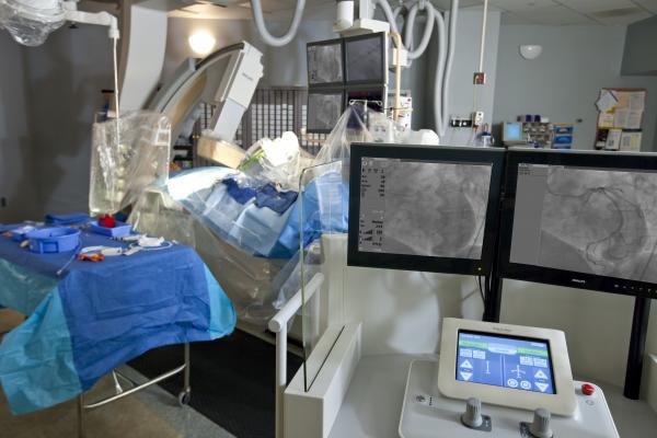
“Historically, the angiography imaging technology emitted more radiation in the past, but the procedures were simpler and did not take as long. Vendors have made improvements and reduced the radiation dose and enable us to still get good images, but we are now doing more complex procedures that take much longer,” said Srihari S. Naidu, M.D., FACC, FSCAI, director, cardiac catheterization laboratory, interventional cardiology fellowship program and hypertrophic cardiomyopathy treatment center at Winthrop University Hospital, Mineola, N.Y. Naidu also is an associate professor of medicine at Stony Brook University, New York (SUNY), and is the chair and founder of the Society for Cardiovascular Angiography and Interventions (SCAI) Emerging Leader Mentorship (ELM) Program. “We do agree to take some risk to help patients, but we are also realizing we need to be more aware of the risks of our radiation exposure. What you can’t see can hurt you. We need to change our behavior to help reduce our exposure,” Naidu said.
He explained that physicians are usually the closest staff to the X-ray source during procedures, so they receive the highest doses. Most of that dose comes from scatter radiation caused by the X-rays defecting off the patient’s tissues and the table. Naidu said most cath lab techs and nurses are usually 5-7 feet away from the X-ray source, which greatly reduces their exposure. Scatter dose rates drop dramatically with just a couple extra feet away from the C-arm and the table, he explained.
Simple Ways to Reduce Dose
Over the past decade, he said angiography systems manufacturers have replaced their image intensifier angiography systems with digital flat panel detectors, which are more sensitive and allow lower-dose imaging. Vendors have also introduced numerous other technology improvements in efforts to lower dose. “This has reduced dose significantly over the past few years,” Naidu said, but added operators can do more to reduce dose in their own labs, possibly reducing dose by 30-40 percent just by changing their habits.
This includes reducing frames rates and better use of collimation. Changing the angulation of the C-arm, positioning the tube in relation to the operator, and reducing the distance between the X-ray source and the patient also can greatly reduce scatter radiation exposure.
Adjusting the table height and where you stand for better ergonomics will also help better distribute the weight of the heavy aprons, Naidu said. During fellowship training, he often tells his fellows not to lean over the table or patient.
During peripheral procedures, digital subtraction angiography (DSA) imaging is used to image vessels, which requires two runs of the imaging system, one using a contrast injection. Naidu explained there is no reason why the operator needs to be in the lab during this imaging sequence and can greatly reduce their exposure by just going into the control room during these imaging runs.
“All of these things have been around forever, but just are not being enforced,” Naidu explained. “We need to be much more methodical about reducing radiation dose.”
Complex Procedures Are Increasing Dose Exposure
The biggest issue facing interventional cardiologists today is that transcatheter procedures have become more complex and take longer than the more basic procedures common several years ago. These newer procedures include opening arteries with chronic total occlusions (CTOs), which can take two or three hours, instead of the easier, straightforward percutaneous coronary interventions (PCIs) that had average times of about 30 minutes.
Peripheral artery disease (PAD) procedures in the legs also tend to be more involved and complex than coronaries. Naidu said peripheral lesions are often long and diffuse, requiring multiple stents and often additional vessel prep with atherectomy or angioplasty. “These things take a long time, unlike focal coronary lesions where you usually only put in one stent and you are done,” Naidu said.
In recent years there has been a rapid increase in transcatheter structural heart procedures for repair of septal defects, left atrial appendage (LAA) occlusion, paravalvular leaks and transcatheter aortic valve replacement (TAVR). These are also much more involved that PCI stent placements.
New Technologies to Reduce Staff Dose in the Cath Lab
Here are five technologies that can help significantly reduce staff radiation exposure from fluoro imaging:
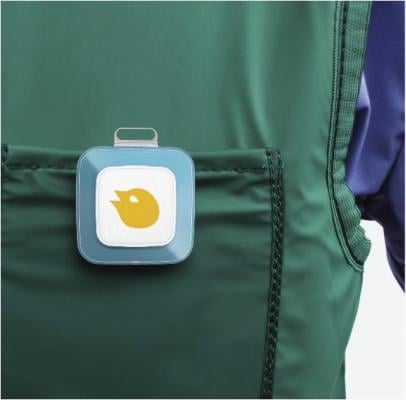
1. Monitoring Dose in Real Time
The traditional radiation badges staff wear are not very helpful because they only alert the badges owner to high levels of radiation well after the fact, often weeks later. One way to make exposure monitoring meaningful is to use a real-time display of radiation levels and show staff members what their exposure is as it is happening. This allows them to react and remove themselves from that radiation field. This is often as simple as taking a step back from the table.
The RaySafe i2 system offers real-time X-ray radiation dose monitoring for cath lab staff, where the detector badges relay live dose data to a display monitor in the cath lab. It is an active dosimetry system that enables effective behavior change by showing physicians and staff their real-time dose during procedures. Teams can all see their personal radiation exposure shown on an overhead screen in the lab and adjust position or make other changes to lower their exposure. The system has visual display, showing colored indications (red, yellow, green) to give each individual user insight about the current dose exposure. The accumulated dose per individual user also is captured and displayed on the touch screen display. The data is stored and can be viewed in a dose dashboard in the system’s dose manager software, which includes total dose history and easily generates reports for staff use and archives.
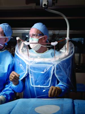
2. Increased Shielding Without the Weight
Tidi Products’s Zero-Gravity Radiation Protection System offers increased radiation protection without weighing down the user. It uses a ceiling suspended gantry system to support a walk-in, lead-lined suit that eliminates the weight of lead aprons being placed on the body of the interventionalist. The gantry allows for smooth movement along the X, Y, Z axis. The lead apron has 1 mm lead equivalency protection from the thyroid to groin, about twice the protection compared to standard lead aprons. It also provides 0.5 mm protection where previously there was none available, and it has front and side protection from direct and scatter radiation. The user can freely engage and disengage without assistance and without breaking sterility.
The Zero-Gravity system has been a big attraction for crowds of cardiologists on the expo floors at both the American College of Cardiology (ACC) and Transcatheter Cardiovascular Therapeutics (TCT) meetings.
3. Robotic Systems to Remove Staff From the Radiation Field
Robotic navigation systems have entered cath and electrophysiology (EP) labs in the last few years as a way to increase precision navigation. These systems also offer the ability for operators to sit at a console away from the table, or in a lead-lined booth without the need to wear lead. This takes the operator out of the radiation field and eliminates back pain issues due to wearing lead protection.
There are three robotic systems currently on the U.S. market that have indications for EP, coronary artery and peripheral artery navigation. Stereotaxis was the first to introduce a robotic system, followed by the Hansen Sensei Robotic System, both designed for long EP procedures. The Corindus CorPath system gained U.S. Food and Drug Administration (FDA) approval for PCI in 2012, followed by an indication for peripheral vessels in March 2016. Hansen’s Magellan is also indicated for peripheral procedures.
Corindus has invested in several studies to collect data on the potential dose savings of its robotic system, which has been found to offer as much as a 95 percent dose reduction. The CorPath has also attracted large crowds at past TCT meetings.
4. Angiography System Advances Reduce Dose
All of the new angiography systems released on the U.S. market over the past few years offer dose-lowering technologies. These advances include improved X-ray tubes, more sensitive detectors, and software to help improve image quality and reduce noise at lower dose settings.
The Philips Allura Clarity is an example of the next-generation system that can help lower standard procedural dose by 50 to 75 percent. The operator can easily adjust frame rates to reduce dose and the system incorporates Philips’ ClarityIQ software to achieve excellent visibility at low X-ray dose levels for patients of all sizes. The software helps correct for motion, reduces noise, auto-enhances the image and corrects pixel shift on cine images.
Toshiba’s Spot Fluoroscopy software for its Infinix-i systems allows clinicians to observe a target region of anatomy using Spot Fluoro’s live fluoroscopy, while viewing the last image hold (LIH) in the surrounding area that has been collimated out of the field of view on the live image to cut dose to both the patient and staff.
The Siemens Artis series incorporates both new X-ray tubes and detectors to lower dose. The new X-ray tube uses flat emitter technology, which enables smaller, square focal spots that the vendor said leads to as much as 70 percent improved visibility of small vessels and improved image quality. The Artis Q.zen combines this X-ray tube with a new crystalline detector that enables patient doses as low as half the standard angiography levels.
5. Lightweight Aprons and Anti-X-ray Hand Cream
BloXR Corp. offers innovative lightweight X-ray shielding aprons for the cath lab composed of bismuth instead of lead or tungsten. Its XPF skirts, vests, aprons, thyroid protectors and caps are approximately half the weight of lead and provide 0.5 mm lead equivalent protection. They are designed to provide clinicians with a lightweight solution to reduce back pain and the limiting mobility of heavy aprons. This protective gear is also machine washable and can be bent, folded and flexed without cracking.
The company also makes an FDA-cleared X-ray attenuating hand cream designed to offer radiation protection when the physician’s hand is in the radiation field. The cream is applied prior to donning gloves, or over a glove with another glove on top, to serve as a lightweight radiation shield. The vendor says it offers a degree of protection for X-ray beams up to 130 kVp.
Related Content on Reducing Radiation in the Interventional Lab:
VIDEO: Technologies and Techniques to Reduce Radiation Dose in the Cardiac Cath Lab — Interview with Akshay Khandelwal, M.D.
VIDEO: Reducing Cath Lab Radiation Dose at Henry Ford Hospital
Read the article, "Helping Interventional Cardiologists Reduce Exposure to Ionizing Radiation."
Read the article "14 Ways to Reduce Radiation Exposure on the Cath Lab."
Watch the VIDEO "Heart Surgeon Shares Effects of Fluoroscopic Radiation Exposure"

 October 24, 2025