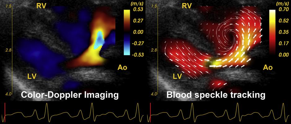
A comparison of color-flow Doppler cardiac ultrasound showing blood flow, and blood speckle tracking revealing a more detailed and complex understanding of the flow. It shows the formation of a vortex that may play a role in future assessments for the efficiency of flow in the heart and vessels. Image from JASE, read more.
In the past several years, a few cardiac ultrasound vendors have developed new ways to image the intricate flow of individual blood cells within the heart, which may soon open new ways to better understand heart diseases. This includes a much better picture of early changes in the blood flow that can lead to heart failure long before symptoms appear, or a better understanding of when to intervene in structural or congenital heart patients.
Currently, only a small percentage of the imaging data that can be acquired in cardiac ultrasound exams is used to create images. Experts in new technology sessions at the American Society of Echocardiography (ASE) meetings over the last decade have predicted that as computing power increases, more of this data will be able to be processed into new, real-time imaging insights.
A few echo vendors have developed blood flow tracking imaging software that is commercially available, but so far there is not enough research to understand how this imaging can be used to help clinical practice.
"Patterns have been observed, but more work needs to be done on the development and validation of quantitative parameters and understanding of how we use them for interventions and clinical practice," explained Partho Sengupta, M.D., in a new technology session at ASE 2021 in June. He is chief of the Division of Cardiology and chief of cardiology, Robert Wood Johnson University Hospital (RWJUH), Barnabas Health and the Henry Rutgers Professor of Cardiology. Until June, Sengupta was the chief of cardiology, director of cardiac imaging, at the West Virginia University Heart and Vascular Institute.
"Blood flow dynamics are extremely intriguing, particularly the symmetric flow across the cardiovascular system. The asymmetry of the blood flow is extremely intriguing and important for a better understanding of cardiac form and function," he said.
Watch examples of this technology from ASE in the VIDEO: Examples of Dynamic Blood Flow Imaging in Cardiac Ultrasound.
Vortices in Blood Flow Add Information on the Efficiency in Fluid Transport in the Heart
By observing blood flow tracking using magnetic resonance imaging (MRI) or cardiac ultrasound, it shows the formation of vortices, which provides information on efficiency for fluid transport in the heart. It is also believed the flow redirection conserves momentum and energy, especially during fast heart rates, Sengupta said. He added that examining blood flow also reveals sheer stresses inside the chambers of the heart and inside blood vessels, which are believed to be involved in the progression of various cardiac diseases, including where coronary plaques form.
"We also just recently realized that abnormalities in sheer stress incites injury and alters cardiac and vasculature form and function, so it may have relevance for cardiac disease formation," he explained.
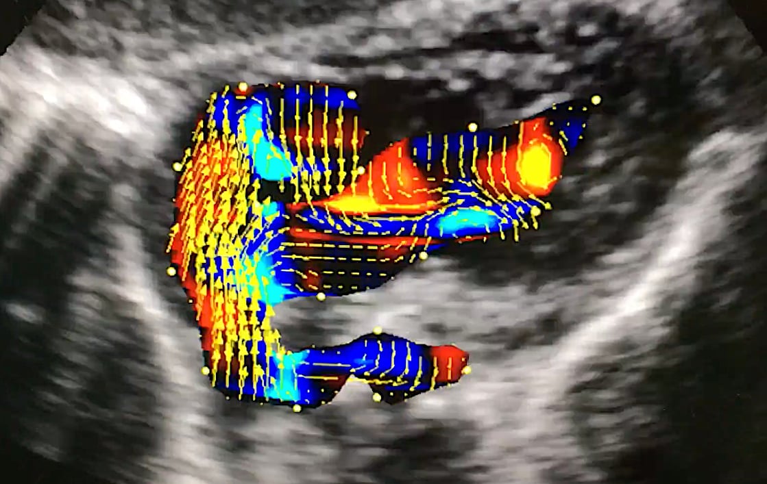
An example of vector flow imaging offered on the Hitachi Lisendo 880 cardiac ultrasound system. Arrows show the direction of the flow and the color coded areas show the velocity. Photo by Dave Fornell at ASE 2018.
Ultrasound Technologies to Image Blood Flow Dynamics
There are three techniques currently used in echocardiography systems to assess blood flow dynamics. These include:
• Particle Imaging Velocimetry, which tracks contrast bubbles moving through the heart.
• Vector Flow Imaging, which images mean velocities of the blood based on color flow Doppler.
• Blood Speckle Tracking, based on pattern matching techniques.
"All three of these technologies currently exist in clinical practice and we are starting to understand the evaluation of blood flow in three dimensions," Sengupta said.
He explained particle imaging velocimetry and blood speckle tracking are both accurate in low flow velocities, but have limits when looking at high velocity flow.[1,2] Vector flow mapping is accurate in high flow velocities, but tends to under-estimate low-flow velocities.[3] For these reasons, Sengupta feels refinement is still needed with all three techniques.
Another hybrid method that has been developed is high-frame-rate echo particle imaging velocimetry, which he said can track echo contrast bubbles at high velocities.[4]
Companies that have developed echo blood flow dynamics imaging on their ultrasound systems include Hitachi, GE Healthcare, Fujifilm, Mindray and BK Medical. All developed vector flow mapping techniques on their ultrasound systems.[5,6,7]
Dynamic Blood Flow Imaging May Offer New Assessment for Congenital Heart Disease
In his presentation, Sengupta showed examples of imaging with normal blood flow through the pulmonary artery with straight lines of flow, and flow in a patient with pulmonary hypertension, where there were lower velocities and vortices that form. He said this appears to show how the inefficient blood flow developed and snowballs into pulmonary hypertension. This type of imaging could be used to detect the early formation of the disease before the patient becomes severely symptomatic.
Another example of a double outlet right ventricle showed large vortices created in the flow, which appear to play a big role in hemodynamic issues in the patient.[2]
"Perturbation of blood flow sequences in congenital heart disease is interesting to observe and may have relevance. It is a very interesting phenomenon, but we do not know yet how to use this in clinical practice. But, we are starting to learn about some of this phenomenon and now being seen, when we otherwise did not see it before," Sengupta explained.
New Types of Cardiac Ultrasound Measurements May Shed New Light on Heart Diseases
Sengupta said most of the work using these imaging techniques show there appear to be opportunities for a better understanding of the heart's form and function based of observing the blood flow. This is especially true in observing vortex formation and perturbation of blood flow sequences in congenital heart disease that may have clinical relevance for better understanding of hemodynamic profiles and the progression and interventions that may help restore the physiology.
Blood flow can be characterized by vortex location, vortex morphology including length, breadth and area, and parameters such as vortex intensity, circulation and formation time. The energy of the vortices also can be measured, including kinetic energy, energy dissipation, energy loss index and energy fluctuation. Pressure gradients and force-flow angles can be measured. Flow transit parameters, such as direct flow and retained flow, also may have application to aid cardiac diagnosis in the future.
"How we use all these parameters in clinical practice we do not yet know," Sengupta explained.
![]()
Examples of GE Healthcare's echo blood flow imaging technique using a combination of high frame rate imaging with blood speckle tracking. It shows how the blood flows within the heart, using arrows to show the direction of flow and color coding showing the velocity. This technique can clearly show vortices created by swirling blood, which are believed to play a role in the early disease development that cause valve issues and heart failure. It also can be used to better assess the hemodynamics of congenital heart defects. The software is being developed for the E95 system. Photos by Dave Fornell at ASE 2019.
Artificial Intelligence Could Play a Key Role in Echo Blood Flow Dynamics Research
Sengupta has been a big advocate for machine learning applications across healthcare, including echocardiography. He said this type of research that involves a large number of parameters where little is known about what the data means is a good example of where artificial intelligence can play a big role in collecting the data and sort it out, making connections with clinically relevant diagnoses. In this case, the answers and data from exams can be fed into the AI, and the machine can sort through it to identify patterns in the dynamic flow imaging that appear to be radiomic markers for the known final diagnosis in a large number of patients.
"One of the approaches we are looking into is to use machine learning techniques to extract all of these parameters and training machine learning algorithms to understand different patient phenotypes and new risk assessment characteristics. This is one of the ways in the future we will be able to extract information and deliver it for for clinical decision support, but more work needs to be done," Sengupta explained.
He said AI also can be used to consider a large array of patient characteristics, including phenotypes, risk characteristics and other data from the the electronic medical record (EMR) to find associations in the imaging data.

An example of several vortices forming in the heart as seen on vector flow imaging on the Hitachi Lisendo 880 cardiac ultrasound system. Arrows show the direction of the flow and the color coded areas show the velocity. Photo by Dave Fornell at ASE 2018.
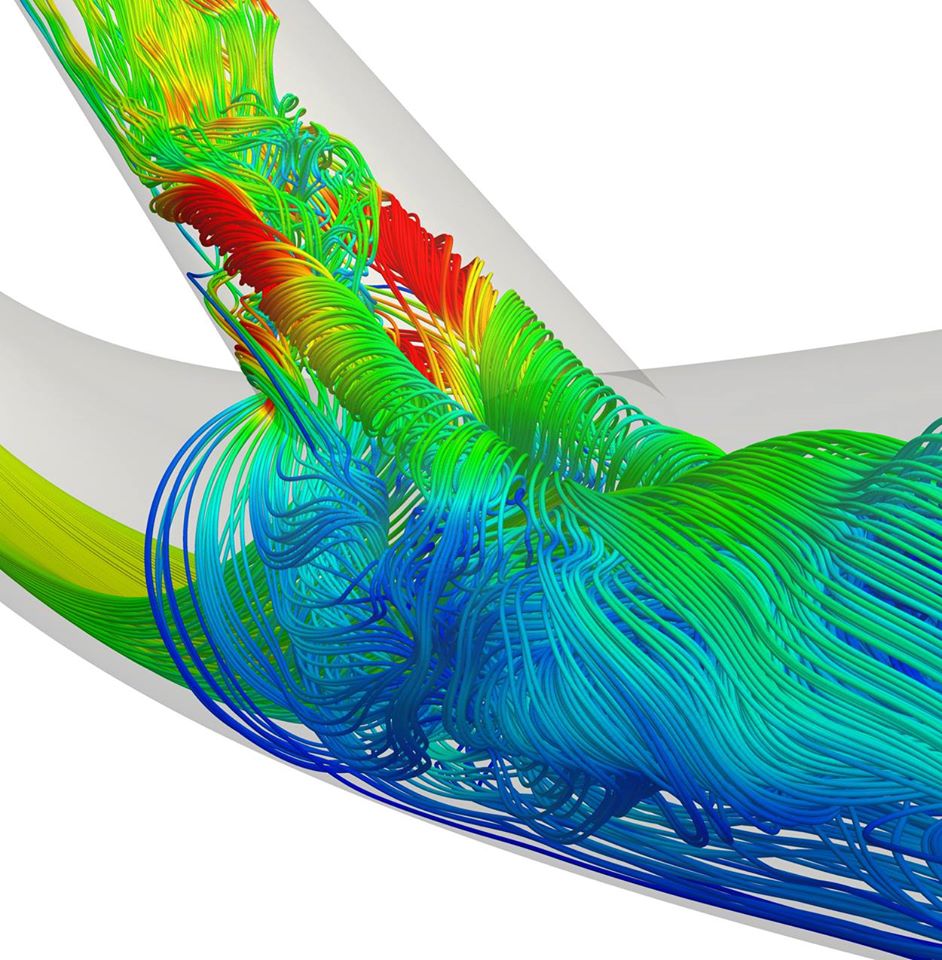
Example of an MRI-derived blood flow fluid dynamics image at the bifurcation of an artery. Research using this type of imaging is expected to shed a better understanding of coronary artery disease formation. Image from the British Cardiology Society.
Related Dynamic Blood Flow Echo Imaging Content:
VIDEO: Examples of Dynamic Blood Flow Imaging in Cardiac Ultrasound
Aurora St. Luke’s Medical Center in Milwaukee Adopts Latest Echocardiography Imaging Software
A Glimpse Into the Future of Cardiac Ultrasound
Analogic Introduces New Premium Cardiac Imaging Software for bk3500 Ultrasound System
Improving Stent Designs With Computational Fluid Dynamics
VIDEO: Editor's Choice of Most Innovative New Cardiac Ultrasound Technology at ASE 2017
References:
3. Tanaka et al. 2021 IEEE International Ultrasonics Symposium. 2021, pp 315-318.
 June 12, 2024