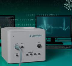
October 28, 2021 — CathVision, a medical technology company developing electrophysiology (EP) solutions in EP recording ...
EP Mapping and Imaging Systems
This channel page contains news on new technology innovations for electrophysiology (EP) mapping and imaging systems used to guide transcatheter cardiac ablation procedures. These systems use mapping catheters that contain electrodes that measure the electrical activity of the cardiac tissue. This is transferred into mapping system software where a 3D model is created of the heart, a color-coded overlay showing the electrical waves generated during each heart beat, the touch points where the tissue was mapped, and showing the location of the catheter inside the heart. Tissue identified as having unhealthy electrical activity that is cause an arrhythmia can then be ablated directly or isolated using an ablation catheter to cause small burns/scar tissue that block electrical signals.
 October 28, 2021
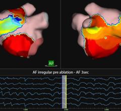
An example of the Acutus Medical AcQMap High Resolution Imaging and Mapping System to guide electrophysiology (EP) cardi ...
October 14, 2021
September 1, 2021 — Stereotaxis and Shanghai Microport EP Medtech Co., Ltd. announced a broad collaboration to advance ...
September 01, 2021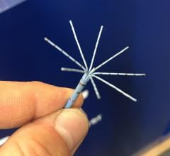
August 3, 2021 – Biosense Webster, the global leader in cardiac arrhythmia treatment and part of Johnson & Johnson ...
August 03, 2021
Jass Brooks, vice president of global strategic marketing, Biosense Webster, explains four over-arching electrophysiolog ...
August 02, 2021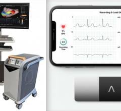
July 27, 2021 — AliveCor Inc., which offers FDA-cleared, smart-phone enabled personal electrocardiogram (ECG) technology ...
July 27, 2021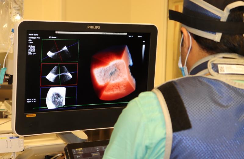
July 21, 2021 — Northwestern Medicine Bluhm Cardiovascular Institute recently became the first cardiovascular program in ...
July 21, 2021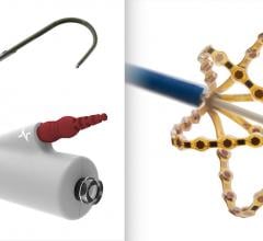
May 25, 2021 — Acutus Medical Inc. announced European CE mark approval for a broad suite of electrophysiology (EP) produ ...
May 25, 2021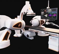
April 7, 2021 — Stereotaxis announced that Broward Health Medical Center is establishing a robotic electrophysiology (EP ...
April 07, 2021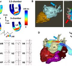
March 23, 2021 — Researchers at Columbia University are using cardiac ultrasound to improve the critical need to ...
March 23, 2021
Feature | Ultrasound Intra-cardiac Echo (ICE) | By Dave Fornell, Editor
Intra-cardiac Echocardiography (ICE) uses catheter-based cardiac ultrasound array to image anatomy and devices inside ...
March 11, 2021
October 8, 2020 – People who suffer from persistent atrial fibrillation in the heart may find relief from a new ...
October 08, 2020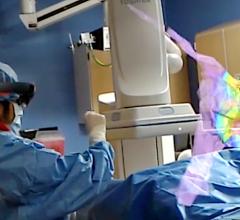
October 1, 2020 — The U.S. Food and Drug Administration (FDA) granted 510(k) clearance for the SentiAR CommandEP system ...
October 01, 2020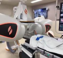
Peter Weiss M.D., MSc, director of ventricular arrhythmia management and robotics, and assistant clinical professor of ...
October 01, 2020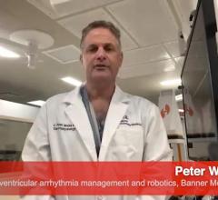
Peter Weiss M.D., MSc, director of ventricular arrhythmia management and robotics, and assistant clinical professor of ...
September 30, 2020© Copyright Wainscot Media. All Rights Reserved.
Subscribe Now