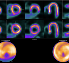
June 28, 2023 — A novel PET perfusion radiotracer, 18F-flurpiridaz, can diagnose coronary artery disease (CAD) in obese ...
PET Imaging
Positron emission tomography (PET) is a nuclear imaging technology (also referred to as molecular imaging) that enables visualization of metabolic processes in the body. The basics of PET imaging is that the technique detects pairs of gamma rays emitted indirectly by a positron-emitting radionuclide (also called radiopharmaceuticals, radionuclides or radiotracer). The tracer is injected into a vein on a biologically active molecule, usually a sugar that is used for cellular energy. PET systems have sensitive detector panels to capture gamma ray emissions from inside the body and use software to plot to triangulate the source of the emissions, creating 3-D computed tomography images of the tracer concentrations within the body.
 June 28, 2023
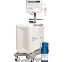
June 26, 2023 — Jubilant DraxImage Inc., dba Jubilant Radiopharma, a wholly-owned subsidiary of Jubilant Pharma Limited ...
June 26, 2023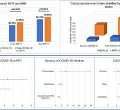 June 09, 2023
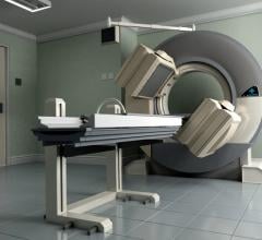 June 05, 2023
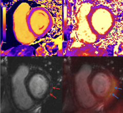
March 10, 2023 — Researchers found evidence of heart muscle inflammation in a small number of patients with acute ...
March 10, 2023![Life Molecular Imaging (LMI) announced that the first patient has been imaged with [18F]florbetaben in the CArdiag Phase 3 clinical study](/sites/default/files/styles/content_feed_medium/public/GettyImages-1413834772.jpg?itok=RX6LEqCg)
February 8, 2023 — Life Molecular Imaging (LMI) announced that the first patient has been imaged with [18F]florbetaben i ...
February 08, 2023
January 5, 2023 — Mouaz H. Al-Mallah, MD, MSc, FASNC, director of the cardiovascular PET program at the Houston ...
January 05, 2023
PET Technology More Effective Than Angiogram at Determining Need for Coronary Stents, Bypass Surgery
December 1, 2022 — A new method for determining whether patients with heart disease need coronary stents or bypass ...
December 01, 2022![Figure 1. Flow chart of machine learning workflow. A machine learning (ML) model for the prediction of vessel-specific ischemia was built, trained, and tuned in the NXT trial (Analysis of Coronary Blood Flow using CT Angiography: Next Steps). The model’s predictive performance was evaluated in the PACIFIC trial (Prospective Comparison of Cardiac Positron Emission Tomography [PET]/Computed Tomography [CT]‚ Single Photon Emission Computed Tomography [SPECT]/CT Perfusion Imaging and CT Coronary Angiography wit](/sites/default/files/styles/content_feed_medium/public/Figure1.jpg?itok=Gz8S08BV)
November 17, 2022 — Cedars-Sinai investigators and colleagues have developed an artificial intelligence (AI) tool that ...
November 17, 2022![Phase III clinical trial of [18F]flurpiridaz PET diagnostic radiopharmaceutical meets co-primary endpoints for detecting Coronary Artery Disease (CAD)](/sites/default/files/styles/content_feed_medium/public/Screen%20Shot%202022-09-13%20at%203.30.13%20PM.png?itok=2w6OoNd6)
September 14, 2022 — GE Healthcare and Lantheus Holdings Inc have announced that the recent Phase III clinical trial of ...
September 14, 2022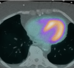
September 7, 2022 — The American Society of Nuclear Cardiology (ASNC) and three partner societies have come together to ...
September 07, 2022
August 25, 2022 — The results of “A Phase 3, Open-label, Multicenter Study of Flurpiridaz (F18) Injection for Positron ...
August 25, 2022
August 12, 2022 — The results of “A Phase 3, Open-label, Multicenter Study of Flurpiridaz (F18) Injection for Positron ...
August 12, 2022
July 11, 2022 – The American Society of Nuclear Cardiology (ASNC) and the Society of Nuclear Medicine and Molecular ...
July 11, 2022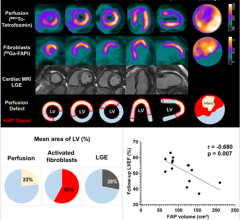
June 15, 2022 — Poor functional outcomes after a heart attack can be predicted with a new PET imaging agent, 68Ga-FAPI ...
June 15, 2022