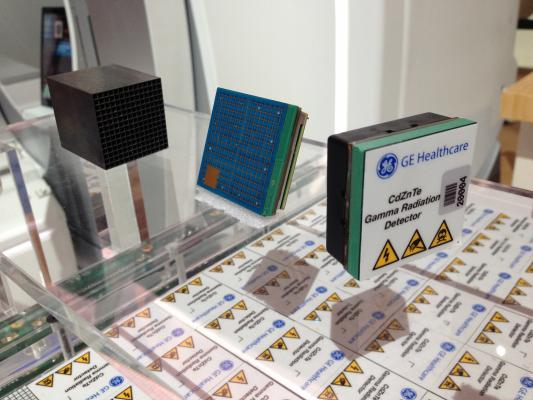
A display of CZT SPECT gamma camera detectors at RSNA 2016. These detectors are more sensitive than those used in older cameras, allowing for faster scans or lower radiation dose.
Cardiac nuclear myocardial perfusion imaging (MPI) has been a mature area of imaging for years, but has recently started a transformation with new technologies, protocols and applications. This is partly due to the availability of technologies, concerns about dose, competition, reimbursement and concerns over isotope availability.
“We have been practicing nuclear cardiology since the late 1970s, so people think of it as a stable and well-established modality, but if you look at the past five years, there has been a tremendous amount of advance in nuclear cardiological techniques,” said Prem Soman, M.D., director of nuclear cardiology at the Heart and Vascular Institute, University of Pittsburgh, and president of the American Society of Nuclear Cardiology (ASNC). “We have a whole new generation of SPECT cameras, we are expanding our imaging applications, we have made great strides in reducing our radiation dose, and PET is becoming more widely used, so I am very excited about the future of cardiac nuclear imaging.”
He said there have been advances in both imaging modalities used for nuclear cardiology, positron emission tomography (PET) and single photon emission computed tomography (SPECT). This includes new, more sensitive SPECT detector technology, new imaging systems and new ways to use the imaging to better quantify perfusion.
Watch the VIDEO “PET vs. SPECT in Nuclear Cardiology and Recent Advances in Technology,” a discussion with Soman at the 2017 ASNC Today meeting.
Newer SPECT Cameras Can Lower Dose or Speed Imaging Time
The traditional analog SPECT gamma cameras use sodium-iodide scintillation crystals, and the new cadmium zinc telluride (CZT) SPECT detectors are based on solid state technology. The new CZT detectors allow for a much smaller footprint for the imaging system, including for office-based imaging or very small imaging suites, Soman explained. The CZT cameras also do not have to rotate around the patient, which makes imaging much easier and efficient than the older technology. The design of the collimators on the newer cameras also offers greater sensitivity. These advantages allow SPECT to be used in new ways, including shorter exams, lower radiation doses or for new techniques like absolute blood flow quantification.
CZT detectors can enable either lower dose or faster imaging speeds. Using normal radioisotope doses, these cameras can image much faster than conventional cameras, reducing scan times or the radioisotope dose exposure, explained Randy Thompson, M.D., attending cardiologist, St. Luke’s Mid-America Heart Institute, Kansas City. However, he said some centers might not be able to take full advantage of the efficiency for faster scans because there are other steps involved in the imaging process that usually cause bottlenecks. “The camera imaging is not really rate limiting to that level,” he explained.
Thompson said St. Luke’s Mid-America Heart Institute uses the radiation dose reduction aspect of the newer cameras. “We have embraced a very low-dose protocol,” he said. “Sometimes it means imaging longer or even repeating images, but radiation dose reduction has been dramatic.”
ASNC recommends half of all patients scanned using SPECT should have a dose of 9 mSv or less of radiation. Thompson said for comparison, 11 mSv is a more standard dose for SPECT. “We now have it down where half of our patients now have a median dose of under 3 mSv,” he said.
To achieve these lower doses, most patients receive stress-only, low-dose exams. “Some of the patients we image this way have a tenth of the dose previously used in the standard approach,” Thompson explained. “If you have enough of those patients in your laboratory, of course the average/median dose comes down a lot. Our average dose in 2009 was about 18 mSv, and now our median dose is around 7.5 mSv, and our mean dose is under 3 mSv.”
However, Thompson said low-dose protocols do not work for all patients. He explained it is difficult to get good image quality with obese patients at lower doses. Some patients might also be better served with faster imaging, including those who cannot hold still for long periods of time because of back problems.
Soman said the old standard dose for SPECT for an average sized patient was about 8-10 millicurie (mCi) of Technetium (Tc-99m) for a resting study and about 24-30 mCi for a stress study. Today, he explained his lab uses a 5 mCi rest and a 15 mCi stress study, resulting in a total body dose of about 6 mSv of radiation. He explained the new ASNC guidelines calling for 9 mSv or less of dose is easily achievable using the new CZT cameras. “You can go down much lower, and there are studies looking at stress-first imaging with only 3-5 mCi of dose. We use that protocol sometimes and the average dose is less than 2 mSv,” he said.
Thompson pointed out lower dose protocols will take time to see wide adoption. The proliferation of the new CZT camera technology into the field also takes time and money, as centers slowly replace older equipment over the course of several years.
“The CZT images do look a little different, and there is a learning curve in reading them,” Soman said. “But, it is not prohibitive and anyone can learn how to read on a CZT system.”
Any centers thinking of replacing older SPECT cameras should consider CZT systems, Soman said. “The efficiencies of the camera and the image quality and the ability to do other things in the future like flow qualification are very good reasons to go with CZT detector technology,” he said.
While the newer CZT scanners offer advantages, Thompson said there are some advantages of the older SPECT scanner technology. This includes the ability to fit extremely obese patients, which may not fit into the smaller confines of the CZT scanners. These cameras also are less expensive. He said a refurbished SPECT scanner may cost half as much as a new CZT scanner, which will allow some smaller centers to still offer nuclear perfusion imaging at an affordable price. However, he said the future of SPECT will be with the newer CZT cameras.
“Many cameras are old and everybody is pressed to buy new equipment, but there are a lot of ways for people to do better quality scans,” said David Wolinsky, M.D., director of nuclear cardiology at Cleveland Clinic Florida and immediate past-president of the ASNC. “Many systems will allow themselves to have software upgrades to do iterative reconstruction. And simply doing better processing will give you better quality images with hopefully less false positives and less need to cath people who end up having normal coronary artieries and do not need a procedure.”
Wolinsky said new CZT SPECT detectors have had a big impact on cardiac nuclear imaging. He said CZT can offer faster cameras, better photon counts and better images. “For older patients you can’t lie down for a long time, you can image them much quicker, and for the younger patients where you don’t want to give them a lot of radiation dose, you can given them a lower dose,” he explained.
Watch the VIDEO “Implementing CZT SPECT Cardiac Protocols to Reduce Radiation Dose,” an interview with Thompson.
Watch the VIDEO “Trends in Nuclear Cardiology Imaging,” an interview with Wolinsky.
PET vs. SPECT Imaging Technologies
“You always want to exercise people on a treadmill if you can and then they can go under a SPECT camera,” Wolinsky said. “You also can get very good adjunct information from an exercise stress test.” With PET, the half-life of the radioisotopes used is very short, so PET imaging only allows for pharmacological stress test imaging, which he said is not ideal. But, for less optimal patients who have issues exercising or who may be high risk, pharmacologic stress is fine to use and they may be better suited to PET.
There are also reimbursement issues regarding PET that play a big role in medical decision-making. He said SPECT is universally reimbursable and it is a lot harder to get a PET scan done. “So, SPECT commands more than 90 percent of the nuclear cardiology market,” Wolinsky said. “So to be able to add better SPECT cameras to allow us to do the same thing we have always done but get better results is really very important.”
For larger institutions and research centers that are imaging larger numbers of high-risk patients, or if they want to image for sarcoid or inflammation, Wolinsky said PET offers a better option.
He said there have been concerns about SPECT radioisotope availability in the past. This prompted new protocols to image using less isotope, which also reduced patient radiation exposure. This includes stress-first or stress-only imaging.
Unlike SPECT, PET does offer some new ways to use nuclear imaging beyond MPI. Soman said this includes a big push with PET to diagnose sarcoidosis, amyloidosis, inflammation and dyssynchrony assessment.
Thompson said some of the newer PET assessments are gaining momentum. He cited a new category III CPT code for a PET myocardial perfusion imaging add-on for absolute quantitation of myocardial blood flow. This goes into effect Jan. 1, 2018. The assessment can help reduce the possibility of false negative exams and improve the accuracy of PET MPI studies. A category III code does not mean the service will be reimbursed, but is seen as a first step. Thompson said it allows providers to speak with payors to ask if the code can be made reimbursable under their insurance plans.
Watch the VIDEO “New CPT Reimbursement Codes for Cardiology,” an interview with Thompson.
Reasons PET Has Not Replaced SPECT
There was a lot of discussion more than 10 years ago when positron emission tomography (PET) first entered the market that it might replace SPECT as the dominant cardiac nuclear imaging technology. However, despite several advantages of PET over SPECT, including improved image quality, PET technology uptake has been very slow. In the intervening years, SPECT technologies have also improved. This includes use of both CZT detectors and iterative reconstruction software to help enhance images and reduce dose.
“PET has some very distinct advantages, but SPECT is much more widely available,” Soman said. “But, there are some big differences in the delivery of the tracer.”
For PET centers that use ammonia as a tracer, the cyclotron that produces the isotope needs to be very close to where the imaging is done due to its very short half-life. With rubidium, an Rb-82 generator is used to create a 75-second half-life tracer in the imaging room. With SPECT, Soman said imaging centers can get a unit dose with much longer half-lives delivered from a remote production facility very easily.
“I think this is one of the reasons why PET technology has taken longer than we expected to implement,” Soman explained. “But, there has been a rapid increase over the past few years for centers doing cardiac PET.”
Combined PET-CT and SPECT-CT Scanners
Much of nuclear imaging is done on smaller SPECT cameras, but the large medical imaging system vendors have pushed the adoption of newer hybrid imaging systems that combine nuclear imaging with a computed tomography (CT) scanner in one gantry. This combination allows for CT attenuation correct on the nuclear images to produce more accurate scans. It also adds CT anatomical image overlays to better visualize the coronary anatomy and better pinpoint where blockages causing perfusion defects are located. Lastly, the CT offers the ability to perform a CT calcium scoring exam of the coronary arteries.
“I think CT adds a lot,” Wolinsky said. “The ability to add calcium scoring is an important piece of information, it adds tremendous value. It is very good for primary prevention and risk identification and adds to the prognostic value of nuclear scans, whether they are PET or SPECT. Also, knowing the calcium score helps you interpret the nuclear scan and helps add prognosis value to the imaging. I think if anybody had a choice, they would want to get a SPECT-CT or a PET-CT.”
However, he said costs for these systems are significant and the people making buying decisions for imaging systems are not the same people as a decade ago. He said some hospital administrators who hold the purse strings today do not always understand the value of nuclear imaging or how an expensive new technology is used to improve patient outcomes. “A hospital really doesn’t care if they make their money doing toenails or treating heart disease, as long as they make money,” Wolinsky explained. “So, we are trying to educate service line managers, hospital administrators and referring physicians about the value nuclear imaging brings to the patient and the service line.”
Basics of Perfusion Imaging and Competition From CT
One criticism of nuclear imaging is found in its industry joke nickname misspelling of “unclear imaging.” Image resolution of SPECT has traditionally shown fuzzy images of the basic shape of the myocardium from the radioactive isotope uptake by the heart muscle and its radiation being detected by cameras outside of the body. This functional imaging shows perfusion defects where there is a lack of radioactive tracer uptake, indicating low or no blood perfusion due to ischemia or an infarct. However, nuclear imaging is unable to accurately show where the exact blockages are located.
CT is traditionally thought of as an anatomical imaging modality, which can produce sharp images of the coronary anatomy, including the ability to visualize blockages inside the arteries. Nuclear imaging has traditionally been used as a follow-up imaging test to determine the severity of blockages on the function of the heart. However, computed tomography (CT) advanced visualization software can now perform perfusion imaging similar to a nuclear scan by mapping iodine contrast levels in the myocardium during the cardiac cycle. Areas of low blood perfusion have lower concentrations of iodine. This software is now offered as part of the post-processing software available with CT scanners. It is also offered by the main third-party CT advanced visualization software vendors. The technology is still being studied in trials to build data to show it is equal to nuclear perfusion imaging. However, cardiac CT advocates say it is only a matter of time before the technology sees wider adoption, especially in the cost-conscious environment of healthcare reform.
Another CT technology that may offer advantages over nuclear is virtual fractional flow reserve (FFR) mapping software. Interventional cardiology uses FFR catheters to invasively measure coronary flow past blockages to determine if the narrowing should be stented or not. FFR-CT can perform a noninvasive FFR assessment for the entire coronary tree at once to show the hemodynamic/functional impact of all blockages. FFR-CT shows a color-coded 3-D reconstruction of the coronary tree that can be rotated on any axis to show the exact source of perfusion defects in the arteries. One software company has had U.S. Food and Drug Administration (FDA) clearance for FFR-CT since November 2014, but there are limitations to the technology — mainly its cost, need to send patient datasets to a supercomputer in California and the time to results, which can take several hours. However, as the technology improves and overcomes these obstacles to adoption and the cost decreases, it may see wider adoption, especially for acute chest pain patients.
Related Content of Nuclear Imaging Advances
Managing Dose in PET and SPECT Myocardial Perfusion Imaging
Advances in Cardiac Nuclear Imaging
PET vs. SPECT - Will PET Dominate Over the Next Decade?
Moving Awareness to Action in Nuclear Medicine Dose
Editor's Choice for Most Innovative Nuclear Imaging Technology Advances at SNMMI 2014
Philips Launches CardioMD IV Cardiac SPECT Solution at ASNC 2017
ASNC and SNMMI Release Joint Document on Diagnosis, Treatment of Cardiac Sarcoidosis
New PET-CT Scan Improves Detection in Rare Cardiac Condition
VIDEO: Clinical Decision Support Requirements for Cardiac Imaging - an interview with Rami Doukky, M.D., system chair, Division of Cardiology, professor of medicine, Cook County Health and Hospitals System, Chicago, at the 2017 American Society of Nuclear Cardiology (ASNC) Today meeting.
New Expert Consensus Outlines Strategies to Improve Myocardial Perfusion Imaging in Women
Study Reveals Low Adoption of IAEA Recommendations for Reduced Nuclear Cardiology Radiation Exposure
Large Nuclear Cardiology Laboratory Slashes Radiation Dose 60 Percent in Eight Years
VIDEO: MACRA's Impact on Cardiology - an interview with Kim A. Williams, Sr., M.D., chief of cardiology at Rush University Medical Center, Chicago and former president of both the American College of Cardiology (ACC) and ASNC, explains the impact of healthcare reform on nuclear perfusion imaging.
University of Missouri Research Reactor Files to Start U.S. Production of Medical Isotopes
New PET Radiotracer Identifies Inflammation in Life-Threatening Atherosclerosis
Automated Radiosynthesis Modules May Improve Cardiac Nuclear Imaging
Societies Call for Focus on Radiation Dose Optimization in Nuclear Cardiology
PET Imaging Visualizes Hard-to-Diagnose Cardiac Amyloidosis
PET/CT Effective in Detecting Calcium Blockages to Assess Heart Attack Risk
New Report Finds U.S. Supply of Critical Medical Isotope Vulnerable
ASNC/SNMMI Release Updated Procedure Standard for PET Nuclear Cardiology Scans
SPECT Systems Comparison Chart
PET/CT Systems Comparison Chart
SPECT-CT Systems Comparison Chart
Comparison charts will require a login, but it is free to register and it only takes a moment.

 January 05, 2023


 imaging program. She spoke on this topic at the 2019 meeting of the American Society Nuclear Cardiology (ASNC) and led a tour with attendees of the PET-CT system at Rush, which was located close to the conference. #ASNC")