Videos

VIDEO: One on One with Hal Wolf, FHIMSS, HIMSS President and CEO

HIMSS | March 05, 2024

Industry trade shows and conferences seem to be making their comeback in 2024. And the Healthcare Information and Management Systems Society (HIMSS) Global Conference and Exhibition seems particularly poised to deliver the best of the best when it comes to digital transformation in both the delivery of healthcare, but also the delivery of a quality experience for those in this demanding, rapidly evolving industry. This month in our ongoing One on One series with industry leaders, we are talking with Hal Wolf, FHIMSS, president and CEO of HIMSS. He offered insights on the society’s new partnership with Informa Markets, key topics being covered at HIMSS24, AI’s impact on the industry, and his thoughts on healthcare sustainability.

Find more HIMSS24 conference coverage here

A New Partnership for Growth

Last August, Informa Markets and HIMSS announced a landmark partnership to propel the growth and evolution of the HIMSS Global Health Conference and Exhibition, recognized as the most influential healthcare technology event of the year, and in North America. It draws 40,000 health professionals, tech leaders, providers and governmental organizations from across the globe. Informa Markets, the world’s largest exhibition organizer, took on management of the HIMSS Exhibition, while HIMSS continues to oversee developing expert content and programming.

Exciting New Features at HIMSS24

At HIMSS2024, with this new collaboration comes new features, including:

Related content:

Find more HIMSS24 conference coverage here

HIMSS Launches Modernized Infrastructure Adoption Model to Support Global Digital Health Transformation

Top Public Policy Experts at HIMSS24 to Address Global AI Landscape and Digital Transformation in Healthcare

VIDEO: Using Maturity Models to Measure Digital Health

VIDEO: Moving Digital Transformation Forward in Healthcare

VIDEO: Key Components to Creating and Implementing AI and Digital Transformation Solutions

VIDEO: The Benefits and Pitfalls of Artificial Intelligence in Healthcare

VIDEO: A Look at Cybersecurity and How Healthcare is at Risk

Conference Coverage

Contrast Media | June 28, 2019

Sharon Mulvagh, M.D., FASE, FACC, FRCPC, professor of medicine, division of cardiology, Dalhousie University, Halifax, Nova Scotia, Canada, and professor emeritus, Mayo Clinic, explains the latest updates to guidelines for the use of cardiovascular ultrasound enhancing imaging agents. She spoke to DAIC at the 2019 American Society of Echocardiography (ASE) meeting.

The changes include a new term for contrast so the agents are not confused with safety issues regarding gadolinium, iodine-based contrast or radioactive nuclear radiotracers. She gives an overview of the 2018 guidelines that are based on a large level of clinical study evidence. Previously, the ASE issued a statement rather than a guideline, because the more clinical evidence was needed for more a more conclusive set of guidelines. 

Read the full guidelines: Clinical Applications of Ultrasonic Enhancing Agents in Echocardiography: 2018 American Society of Echocardiography Guidelines Update.

Find more news and videos from ASE 2019.


Artificial Intelligence | June 28, 2019

This is a quick example of how artificial intelligence (AI) is being integrated on the back end of cardiac ultrasound systems to help automate and speed workflows. This video segment is from the expo floor of the 2019  American Society of Echocardiography (ASE) meeting. It shows AI-based AFI automation features incorporated into the GE Vivid E95 echo system. 

Read a blog about how AI will help advance cardiac ultrasound imaging, Combatting the No. 1 Cause of Death With the Help of Artificial Intelligence and Advanced Technology.

Find more ASE 2019 coverage


Heart Failure | June 28, 2019

Federico Asch, M.D., FASE, director of cardiac imaging research and director of the cardiovascular imaging lab, MedStar Health Research Institute, Washington, D.C., explains the importance of the COAPT Trial and the cardiac ultrasound findings from the study. It was presented as a late-breaking study at the American College of Cardiology (ACC) 2019 meeting and the sub-study was discussed in several sessions at the 2019 American Society of Echocardiography (ASE) conference.

The trial showed the Abbott MitraClip can be used to help large numbers of patients with heart failure who experience increasing levels of mitral valve regurgitation due to the enlargement of their hearts. 

Read more about the COAPT Trial

VIDEO: MitraClip to Treat Heart Failure - Results of the COAPT Trial — Interview with William Abraham M.D. 

VIDEO: Impact of the COAPT Trial on Heart Failure Patients With Functional Mitral Regurgitation — Interview with Andreas Brieke, M.D.

FDA Approves MitraClip for Use in Heart Failure Patients With Functional Mitral Regurgitation

360 View of the TEE Echo Workstation During a MitraClip Procedure


Cardiovascular Ultrasound | June 27, 2019

Judy Hung, M.D., director of echocardiography, Division of Cardiology, Massachusetts General Hospital, Boston, explains some of the changes in recent guidelines for cardiac ultrasound assessments of aortic and mitral valves. She spoke at the 2019 American Society of Echocardiography (ASE) meeting.


Links to ASE guidelines:

   • Recommendations for Noninvasive Evaluation of Native Valvular Regurgitation

   • Recommendations on the Echocardiographic Assessment of Aortic Valve Stenosis: A Focused Update from the European Association of Cardiovascular Imaging and the American Society of Echocardiography


Find more ASE 2019 coverage


Virtual and Augmented Reality | June 27, 2019

Roberto Lang, M.D., director of cardiac imaging at the University of Chicago, has been working with TomTec for the past three years on a project to use virtual reality (VR) to edit and view 3-D cardiac ultrasound and CT scans. He spoke at a couple sessions this week at the 2019 American Society Of Echocardiography (ASE) meeting, where doctors were able to view the session using VR headsets. The hands-on demonstration of this technology in the TomTec booth at ASE 2018 and 2019 was one of the most popular exhibits with attendees both years.

Read more about this technology in Top Technology Trends in Echocardiography at ASE 2018

See examples of VR technology for echo in the  VIDEO: Editor’s Choice of the Most Innovative Echo Technology at ASE 2018

Here is a video example of this technology being demonstrated at ASE19.


Cath Lab | June 26, 2019

Thomas Porter, M.D., FASE, the Theordore F. Hubbard Distinguished Chair of Cardiology and a professor of medicine at the University of Nebraska Medical Center, explains a new study on how cardiac ultrasound imaging combined with bubble contrast was able to break down blood clots and restore blood flow to blocked vessels in STEMI heart attack patients. He gave an update on this new potential therapeutic direction for ultrasound at the 2019 American Society Of Echocardiography (ASE) meeting.
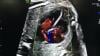
The Brazilian study used this approach in about 100 ST-elevated myocardial infarction (STEMI) patients and was recently published in the Journal of the American College of Cardiology (JACC). Porter said about 50 percent of patients had their ST resolved by the time they made it to the cath lab for percutaneous coronary intervention (PCI). Two more studies in Amsterdam and Pittsburgh are now starting to see if this technique can be used while transporting patients in an ambulance to further improve heart attack outcomes and to reduce infarct size.

Porter also explains recent research that shows ultrasound pulses promote ATP and nitric acid production, which have a lasting beneficial effect on small vessel perfusion in the area treated and down stream for hours after the use of the transducer. 

Watch the VIDEO: Therapeutic Applications for Contrast Ultrasound — another  interview with Porter. 


Wearables | June 21, 2019

Jacques Kpodonu, M.D., FACC, cardiac surgeon, Beth Israel Deaconess Medical Center, and professor at Harvard Medical School, explains how medical devices and wearables that interface with smartphones and apps might be used to eliminate healthcare disparities in rural areas of the developed world and help raise the level of care in the developing world. He spoke at the 2019 AI-Med Cardiology conference. 


Related Smartphone and Wearable Content:

VIDEO: Use of Wearable Medical Devices for Cardiac Rehabilitation — Interview with Robert Klempfner, M.D.

VIDEO: Use of Wearables to Track Electrophysiology Patients — Interview with Khaldoun Tarakji, M.D.

Smartphones Used to Successfully Screen More than 60,000 for Atrial Fibrillation

VIDEO: The Future of Wearables in Healthcare — Interview with Karl Poterack, M.D.

VIDEO: Artificial Intelligence Applications for Cardiology — Interview with Anthony Chang, M.D.
 

Atrial Fibrillation | June 21, 2019

Sanjaya Gupta, M.D., electrophysiologist, St. Luke's Mid America Heart Institute, and assistant professor, University of Missouri–Kansas City School of Medicine, explains how his center developed an artificial intelligence (AI) application to automatically risk stratify atrial fibrillation (AFib) patients. The Epic-based app stratifies patients into those who should be placed on anticoagulation and those who are candidates for left atrial appendage (LAA) occlusion. He spoke at the 2019 AI-Med Cardiology conference

His center hopes to develop similar guidelines based AI apps for other types of cardiac risk scoring. Gupta said he is looking for other centers to partner with to co-develop and test these AI apps.    


Related Cardiology AI Content:

VIDEO: Overview of Artificial Intelligence and its Use in Cardiology — Interview with Anthony Chang, M.D.

VIDEO: ACC Efforts to Advance Evidence-based Implementation of AI in Cardiovascular Care — Interview with John Rumsfeld, M.D.

VIDEO: Artificial Intelligence Applications for Cardiology — Interview with Anthony Chang, M.D.

PODCAST: Fitting Artificial Intelligence Into Cardiology — Interview with Anthony Chang, M.D.

VIDEO: How Hospitals Should Prepare for Artificial Intelligence Implementation — Interview with Paul Chang, M.D.

Technology Report: Artificial Intelligence 

Artificial Intelligence | June 20, 2019

John Rumsfeld, M.D., Ph.D., FACC, American College Cardiology (ACC) chief innovation officer, and professor of medicine at the University of Colorado School of Medicine, explains how the ACC is working with artificial intelligence (AI) vendors to directly impact cardiac care. He said there is a tremendous amount of investment in and hype surrounding AI in healthcare, but to date there has been very little of this has translated in to changes in the way cardiology care is delivered. He outlines several areas to successfully apply AI to improve cardiovascular care and outcomes. He also discussed the current ACC efforts to advance evidence-based implementation of AI in cardiac care including applications for the NCDR.

He spoke at the 2019 Cardiology AI-Med conference

Watch the related VIDEO: Overview of Artificial Intelligence and its Use in Cardiology, an interview with Anthony Chang, M.D., chief artificial intelligence officer, Children’s Hospital of Orange County (CHOC), and founder of AIMed.  


Artificial Intelligence | June 18, 2019

Anthony Chang, M.D., chief artificial intelligence officer, Children’s Hospital of Orange County (CHOC), and founder, AIMed, explains the basic principles of artificial intelligence (AI) in medicine. He outlines some basic AI definitions, potential programming biases and use cases. He also explains the need for the Cardiology AI-Med conference, which held its inaugural meeting in June 2019 in Chicago.


Related AI Content:

Link to all the recorded AI-Med Cardiology conference sessions

VIDEO: Artificial Intelligence Applications for Cardiology — Interview with Anthony Chang, M.D.

PODCAST: Fitting Artificial Intelligence Into Cardiology — Interview with Anthony Chang, M.D.

VIDEO: How Hospitals Should Prepare for Artificial Intelligence Implementation — Interview with Paul Chang, M.D.

Technology Report: Artificial Intelligence 

Heart Valve Technology | May 20, 2019

A demonstration of how to calculate the neo-left ventricular outflow tract (neo-LVOT) on CT imaging for a transcatheter mitral valve replacement using Circle Imaging's advanced visualization software. The demonstration looks at the use of an Edward's Sapien valve being implanted for a mitral valve-in-valve procedure. The overhang of the Sapien can block the LVOT blood flow, which can be catastrophic for the patient. So, assessment of the neo-LVOT in a simulation of the implant is required prior to the procedure to find the ideal landing zone and assess if the patient's anatomy is compatible with this technique.  

Watch the related VIDEO: The Importance of the Neo-LVOT in Transcatheter Mitral Valve Replacement — an interview with Dee Dee Wang, M.D., director of structural heart imaging, Henry Ford Hospital. 

This clip was recorded on the expo floor at the 2018 Society of Cardiovascular Computed Tomography (SCCT).


Cardiovascular Ultrasound | May 16, 2019

This is an example of how the heart's left atrial appendage (LAA) can be evaluated for thrombus and possible transcatheter occlusion using a new cardiac ultrasound lighting technology called TrueVue. It is a movable virtual light source that can interact with the echocardiography images to show photorealistic, virtual surgical views of the cardiac anatomy. The light source can be moved anywhere in the image, including behind structures to backlight them. The technology is offered on the Philips Healthcare Epiq CVx cardiovascular ultrasound system. It was shown for the first time in the U.S. at the American Society of Echocardiography (ASE) 2018 meeting.

See another VIDEO example of the photo-realistic lighting technology showing a transcatheter ASD closure with two Amplatzer occluders.


Stroke | May 16, 2019

This is an example of a carotid artery reporting module from Change Healthcare at 2018 Radiological Society of North America (RSNA) annual meeting. It shows how the PACS can bring in ultrasound imaging of the carotid artery and the graphical report can be modified to match the patient anatomy. The text and modifications made to the vessel tree convert into text to help auto-fill fields in the written report to help speed workflow. The vessel tree is similar to cath lab reporting systems that use a similar model of the coronaries that can be modified and helps auto complete the cath report.


Related Content:

VIDEO: What to Look for in PACS Workflow Efficiency

6 Key Health Information Technology Trends at HIMSS 2019

The Building Blocks of Enterprise Imaging

Technology Report: Enterprise Imaging

Find more RSNA 2018 coverage.


Information Technology | April 17, 2019

With Intellispace Enterprise Edition as the foundation, Philips Healthcare is connecting facilities and service areas within enterprises, while developing standards-based interoperability that preserves customers' investments and best of breed systems. 

Artificial Intelligence | April 17, 2019

Paul Chang, M.D., professor of radiology, vice chair of radiology informatics and medical director for enterprise imaging, University of Chicago, explains some of the issues with artificial intelligence (AI) and how hospitals can better prepare for its eventual implementation across the field medicine. A key takeaway is that hospitals need an infrastructure and roadway for AI and deep-learning algorithms to operate. Chang said most health systems will not invest directly in AI, but will invest in analytics, which Chang said uses much of the same infrastructure required by AI.

Chang spoke on this topic at an AIMed breakfast briefing seminar in Chicago April 9, 2019. Listen to a webcast of this hour and 15 minute talk.


Wearables | March 26, 2019

Khaldoun Tarakji, M.D., MPH, staff physician in the Section of Electrophysiology and Pacing in the Robert and Suzanne Tomsich Department of Cardiovascular Medicine, Sydell and Arnold Miller Family Heart and Vascular Institute at the Cleveland Clinic, explains how wearable devices and smart phone apps can be used to aid electrophysiologists in patient care. He said the devices offer a constant remote monitoring of patient heart data, which can be helpful in diagnosing various types of arrhythmias and cardiac conditions. However, the main issue is how to sort through the large volumes of data and to figure out what the clinical value of some of this consumer data is through studies.  He spoke at the American College of Cardiology (ACC) 2019 meeting.


Other Cardiac Wearable Content:

VIDEO: Use of Wearable Medical Devices for Cardiac Rehabilitation — Interview with Robert Klempfner, M.D.

VIDEO: The Future of Wearables in Healthcare — Interview with Karl Poterack, M.D.


Cardiovascular Business | March 26, 2019

William Pinsky, M.D., a pediatric cardiologist and CEO of the Educational Commission for Foreign Medical Graduates (ECFMG), and Mandeep Mehra, M.D., medical director of the Brigham and Women’s Hospital (BWH) Heart and Vascular Center and professor of medicine at Harvard Medical School, explain the U.S. doctor shortage and how foreign doctors help fill the gap.

According to 2017 data provided by the Association of American Medical Colleges (AAMC), 40 percent of interventional cardiologists, 30 percent of cardiovascular disease specialists, and 26 percent of pediatric cardiologists in the United States are international medical graduates (IMGs). However, as the physician shortage continues to impact primary care doctors, psychiatrists, OB/GYNs, among others, the U.S. also expects to see a shortage of cardiologists within the next 10 years, according to a spotlight cardiology study issued by the professional services firm PYA, which specialized in healthcare consulting.

The interview was shot at the American College of Cardiology (ACC) 2019 meeting.


Cardio-oncology | March 22, 2019

Magid Awadalla, MBBS, is an advanced cardiac imaging research fellow at Massachusetts General Hospital. He has been involved in an imaging study of cardiac changes from photon radiotherapy in breast cancer patients using serial cardiac magnetic resonance imaging (MRI). The radiotherapy beams used to treat breast cancer pass close to the neighboring heart, which can cause cardiac cell damage leading to issues like heart failure later on. He spoke on the topic of cardio-oncology at the American College of Cardiology (ACC) 2019 meeting.

Nuclear Imaging | March 22, 2019

Raza Alvi, M.D., a research fellow in radiology at Massachusetts General Hospital, has been involved in a study of a positron-emission tomography (PET) FDG radiotracer agent to image sarcoidosis. The inflammatory disease affects multiple organs and usually include abnormal masses or nodules (granulomas) consisting of inflamed tissues that can form in the heart. Alvi presented on this topic at American College of Cardiology (ACC) 2019 meeting


Related Cardiac Sarcoidosis Content:

ASNC and SNMMI Release Joint Document on Diagnosis, Treatment of Cardiac Sarcoidosis

New PET-CT Scan Improves Detection in Rare Cardiac Condition

25 Most Impactful Nuclear Cardiology Articles

Recent Advances in Cardiac Nuclear Imaging Technology

EP Lab | March 21, 2019

Khaldoun Tarakji, M.D., MPH, associate section head, section of electrophysiology and pacing in the Robert and Suzanne Tomsich Department of Cardiovascular Medicine, Sydell and Arnold Miller Family Heart and Vascular Institute at the Cleveland Clinic. He presented the WRAP-IT late-breaking trial at the American College of Cardiology (ACC) 2019 meeting. The trial looked at the use of an antibacterial envelope for pacemakers and ICDs to reduce infection risk. 

Read the ACC.19 article Medtronic Tyrx Envelope Significantly Reduces Major Infections in Cardiac Implantable Device Patients.


Rlated EP video From ACC.19:

VIDEO: Key Trends in Electrophysiology — Apple Watch to Detect AF and the CABANA Trial — Interview with Christine Albert, M.D.


Wearables | March 08, 2019

Karl Poterack, M.D., medical director, applied clinical informatics, Mayo Clinic, explains the role wearable devices will play in healthcare. He presented in several sessions at the 2019 Healthcare Information Management and Systems Society at (HIMSS) conference.

Poterack said there is a brewing tsunami of data in wearable technologies that healthcare systems will have to figure out how to integrate in the coming years. He said the key issue with wearable data is that there needs to be outcomes data showing the value of how many steps a patient accumulates, changes in heart rate over time, or blood pressure changes in patients with specific aliments. Without this , he said there is limited value in the information. 

Watch the related VIDEO: Use of Wearable Medical Devices for Cardiac Rehabilitation.

Look through a photo gallery of other new technologies at HIMSS19. 

Find news and videos from HIMSS 2019.

Advanced Visualization | March 05, 2019

Augmented reality (AR) and virtual reality (VR) are starting to be adopted for physician training, patient education about their planned procedures, treatment planning and it is expected to be used as a procedure guidance tool in the near future. This example of AR displayed at the Healthcare Information Management and Systems Society (HIMSS) 2019 meetingwas among the most innovative because it allows users to "feel" the 3-D hologram of the heart. Developed by the company SoftServe., the “Touch My Heart” work-in-progress technology allows anyone wearing an AR headset to see and interact with the heart and get a touch sensation when they reach into the virtual tissue. A pad below the image is composed of dozens of ultrasound transducers that emit sound waves in the shape of the heart so users feel touch sensations when interacting with the virtual tissue.

Read the article "Virtual Reality Boosts Revenues and Patient Understanding,"

Look through a photo gallery of other new technologies at HIMSS19. 

Find news and videos from HIMSS 2019.


ECG | March 05, 2019

This is a quick demo of the Schiller Cardiovit FT-1 electrocardiograph (ECG) system displayed at the Healthcare Information Management and Systems Society (HIMSS) 2019 meeting. It has a 3-D rendering of a patient showing where each lead needs to be placed. The user can rotate the images on the touch screen to see where the leads go and can easily identify where any issues are when the system automatically alerts them about misplaced leads. The goal is to improve and speed ECGs using a better form of visualization than the traditional black and white 2-D pictures. The system changes the lead place placements of the body rending based on the type of exam being performed using a drop down menu.

Look through a photo gallery of other new technologies at HIMSS19. 

Find news and videos from HIMSS 2019.

Artificial Intelligence | March 04, 2019

Anthony Chang, M.D., chief intelligence and innovation officer, Children's Hospital of Orange County (CHOC), and medical director of the Sharon Disney Lund Medical Intelligence and Innovation Institute. He is expert in artificial intelligence (AI). He spoke in several sessions at Healthcare Information and Management Systems Society (HIMSS) 2019 meeting on the integration of AI in healthcare. 

He said AI will play a big roll in imaging assessments of adult congenital heart disease to help relieve the burden on the small number of congenital cardiologists. 

Chang also explained there is a tsunami of data about to wash over healthcare as wearable devices begin to be integrated into patient care. AI will play a key role in sorting through all this data by monitoring the information to identify trends or disease markers and alert clinicians and the patient.

He was a keynote speaker at HIMSS19 with his session "Synergies Between Man and Machine — Future AI apps can be directed to help mitigate physician burnout by decreasing the EHR burden, improving medical education, and automating quality improvement."

Chang is head of the artificial intelligence organization AIMed, which hosts educational sessions and an annual meeting on AI applications in medicine.

Listen to Chang in the PODCAST: Fitting Artificial Intelligence Into Cardiology.
 

Read the article 6 Key Health Information Technology Trends at HIMSS 2019.

Look through a photo gallery of other new technologies at HIMSS19. 

Find news and videos from HIMSS 2019.

Wearables | February 28, 2019

Robert Klempfner, M.D., director of the Cardiovascular Prevention Institute, Sheba Medical Center, Israel,  discusses his center's use of wearable devices to manage a remote cardiac rehabilitation program. He spoke on the topic at 2019 Healthcare Information Management and Systems Society at (HIMSS) conference.   

Sheba Medical Center in Israel has adopted an app that interfaces with various wearables and Bluetooth-enabled patient monitoring devices to create remote cardiac rehabilitation and heart failure programs. It now remotely monitors hundreds of patients and does not require them to come to the hospital for sessions, which has helped increase patient satisfaction and aided in increasing physical activity compliance. The app is able to transfer device data to an EMR so progress and tasks assigned to patients can be monitored without the need for them to come into the hospital for sessions. Klempfner recently wrote a paper on this topic

Watch the related VIDEO: The Future of Wearables in Healthcare 

VIDEO: Mobile App Links Wearable Data to Electronic Medical Records — The Datos software used by Sheba

Look through a photo gallery of other new technologies at HIMSS19. 

Find news and videos from HIMSS 2019.


Wearables | February 28, 2019

How wearable devices will play a role in healthcare was a big topic at the Healthcare Information Management and Systems Society (HIMSS) 2019 meeting. The biggest question is how to attached the data from consumer devices into a usable format for clinicians that interfaces with the electronic medical record (EMR). A good example of how wearables are being integrated in clinical care was demonstrated by the company Datos. It offers software that can integrate data from a wide variety of wearable devices from several makers into a mobile app, It can transfer the information to an EMR. The app also offers two way communication between the patient and the doctor’s office. It can prompt patients with a list of things they need to do each day to reach physician specified goals and displays analytics on a patient’s health data, including charts and graphs.

The system is used by Sheba Medical Center in Israel for a remote cardiac rehabilitation program. It now remotely monitors hundreds of patients and does not require them to come to the hospital for sessions, which has helped increase patient satisfaction and aided in increasing physical activity compliance. Watch the VIDEO: Use of Wearable Medical Devices for Cardiac Rehabilitation — an interview with Robert Klempfner, M.D., director of the Cardiovascular Prevention Institute, Sheba Medical Center.
 

Look through a photo gallery of other new technologies at HIMSS19. 

Find news and videos from HIMSS 2019.

EP Lab | February 27, 2019

This is a virtual heart with the same electrophysiology characteristics as the real patient being developed to help optimize cardiac resynchronization therapy (CRT) lead placement. CRT currently has a 30 percent nonresponder rate, which is mainly due to the placement of leads. This model allows virtual placement of the leads In various locations to test response prior to the implantation procedure. The green dot shows the location of the virtual lead. It was unveiled at the Healthcare Information Management and Systems Society (HIMSS) 2019 annual meeting in February by Siemens. This "digital twin" technology is in development and will be able to create virtual, digital organs from a patient’s ECG, MRI scan and other clinical data. Siemens said the technology also might have applications for testing virtual ablations strategies to save procedure time when the patient is in the EP lab

Read more about the digital twin technology.

Look through a photo gallery of other new technologies at HIMSS19. 

Find news and videos from HIMSS 2019.


Enterprise Imaging | February 27, 2019

Steve Holloway, principal analyst and company director for the healthcare market research firm Signify Research, explains the key trends he is seeing in enterprise imaging systems. He spoke to ITN at the 2019 Healthcare Information Management and Systems Society at (HIMSS) conference.  

Additional HIMSS 2019 coverage.

Look through a photo gallery of new technologies highlighted at HIMSS 2019.

Watch the RSNA 2018 VIDEO: Technology Report — Enterprise Imaging

Artificial Intelligence | January 28, 2019

DAIC Editor Dave Fornell takes a tour of some of the most interesting new artificial intelligence (AI) imaging technologies on the expo floor of 2018 Radiological Society of North America (RSNA) annual meeting. Several of the new AI apps aid in faster iidentification of stroke, help in interventional vascular guidance for live embolizations, and speed workflow in medical imaging and reporting systems.

Watch the related VIDEO: Editor's Choice of the Most Innovative New Imaging Technologies at RSNA 2018


Related AI Imaging Technology Content:

New Cardiac Imaging Technologies Unveiled at RSNA 2018

VIDEO: Overview of Artificial Intelligence and its Use in Cardiology — Interview with Anthony Chang, M.D.

VIDEO: How Hospitals Should Prepare for Artificial Intelligence Implementation — Interview with Paul Chang, M.D.

VIDEO: Managing a Multi-site Radiology Practice With AI-based Workflow — Interview with Andrew Deutsch, M.D.

VIDEO: Technology Report: Artificial Intelligence

VIDEO: RSNA Post-game Report on Artificial Intelligence — ITN editors Dave Fornell and Greg Freiherr discuss the AI trends they saw at RSNA 2018

VIDEO: RSNA President Vijay Rao Says Artificial Intelligence is Hottest Tech Advancement in Radiology 

How Artificial Intelligence Will Change Medical Imaging


How to Market Healthcare Artificial Intelligence Software

Increasing Presence of AI at RSNA Reflects Emphasis on Efficiency

VIDEO: AI, Analytics and Informatics: The Future is Here — Interview with RSNA 2017 keynote speaker Michael Recht, M.D.

VIDEO: A Walk Through the RSNA 2018 Machine Learning Showcase

Selecting an AI Marketplace for Radiology: Key Considerations for Healthcare Providers


Increasing Presence of AI at RSNA Reflects Emphasis on Efficiency

Technology Report: Artificial Intelligence at RSNA 2018

RSNA 2018 Key Takeaways from the Expo Floor

VIDEO: How iCad Uses AI to Speed Breast Tomosynthesis

VIDEO: How Imalogix Uses AI to Boost Performance


VIDEO: Collaboration as a Catalyst for AI Innovation

PODCAST: How to Make Artificial Intelligence a Success in Medicine

VIDEO: Example of AI-assisted Oncology Patient Record Dashboard


Cardiac Imaging | January 26, 2019

DAIC Editor Dave Fornell takes a tour of some of the most interesting new medical imaging technologies displayed on the expo floor at the 2018 Radiological Society of North America (RSNA) meeting. The video includes new technologies for fetal ultrasound, CT, MRI, mobile DR X-ray, a new generation of fluoroscopy systems, MRI contrast mapping to better identify tumors, and a new technique to create moving X-ray images from standard digital radiography (DR) imaging.

Watch the related VIDEO: Editor’s Choice of the Most Innovative New Artificial Intelligence Technologies at RSNA 2018. This includes a tour of some of the recently FDA-cleared AI technologies for medical imaging at RSNA 2018. 


Related Content:

New Cardiac Imaging Technologies Unveiled at RSNA 2018

VIDEO: Overview of Artificial Intelligence and its Use in Cardiology — Interview with Anthony Chang, M.D.

VIDEO: How Hospitals Should Prepare for Artificial Intelligence Implementation — Interview with Paul Chang, M.D.

Echocardiography Trends at ASE 2019

7 Hot Topics in Cardiac CT Imaging


December 12, 2018

This is a walk around of the new Spectrum Dynamics Veriton SPECT-CT nuclear imaging system introduced at the 2018 Radiological Society of North America (RSNA) meeting.

This is a walk around of an innovative new SPECT-CT nuclear imaging system shown at the Radiological Society Of North America (RSNA) 2018 meeting this week. It's CT system with comes in 16, 64 or 128 slice configurations. It has 12 SPECT detector robotic arms that automatically move toward the patient and use a sensor to stop a few millimeters from the skin to optimize photon counts and SPECT image quality. It also uses more sensitive CZT digital detectors, which allows either faster scan times, or use of only half the radiotracer dose of analog detector scans.

Read the article “Nuclear Imaging Moves Toward Digital Detector Technology.” 

Read the article "Spectrum Dynamics Sues GE for Theft, Misappropriation of Trade Secrets and Unfair Competition."


Cardiovascular Ultrasound | December 12, 2018
This is an example of the new Fetal HQ heart and vascular software from GE Healthcare for fetal ultrasound. The software, for the Voluson E10, helps evaluate the fetal heart shape, size and contractibility. A feature called Radiant Flow shows the blood flow in a 3-D view. It can also help show slow-flow blood, such as neuro-vascular circulation. This was shown for the first time at the 2018 Radiological Society of North America (RSNA) meeting.


Advanced Visualization | December 12, 2018

This is an example of the FDA-cleared OpenSight augmented reality (AR) system for surgical planning from NovaRad at the Radiological Society Of North America (RSNA) 2018 meeting. It uses a HoloLens headset to register an overlay of the patient’s MR or CT scan in the patient. The operator can use hand movements to slice through and manipulate the images.

The vendor gained an FDA indication for AR to be used in surgical planning in 2018. NovaRad is working with the FDA for a second indication for use of the AR in the operating room during procedures.

This video is jerky, slightly misaligned and the hand movements did not always respond because itwas shot with an iPhone inside the visor. The image quality and hand movements are much better when actually wearing it on your head and aligned for the specific user.

Read more about this technology

Cybersecurity | December 12, 2018

Anton S. Becker, M.D., radiology resident at the University Hospital of Zurich, Switzerland, discusses the long-term risks of cyberattacks on medical imaging data at the 2018 Radiological Society of North America (RSNA) annual meeting.

Subscribe Now