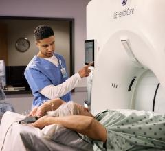
Siemens Go.Top cardiovascular edition CT scanner with its detachable tablets used by the tech to control the scanner so they can stay at the patient's side longer. (Photo by Dave Fornell)
There were several interesting new trends in cardiovascular computed tomography (CT) imaging at the 2019 Society of Cardiovascular Computed Tomography (SCCT) meeting in July. Key topics at this year's meeting included integration of artificial intelligence (AI) into CT systems, the integration of CT calcium scoring into the 2018 American Heart Association (AHA) cholesterol management guidelines, structural heart assessments for transcatheter valves and left atrial appendage (LAA) occlusion, and first-time partner sessions with other cardiac societies. This was according to SCCT President Ron Blankstein, M.D., director of cardiac computed tomography, Brigham and Women's Hospital, and associate professor of medicine and radiology, Harvard Medical School.
"The new guidelines have certainly helped propel the use of calcium scoring forward and it is a big highlight at this meeting. We have seen the calcium scoring sessions here packed," Blankstein said. "There also have been a lot of new data presented on the use of artificial intelligence in CT and how we can use it for things like automated plaque quantification."
![]() Watch an interview with Blankstein in the VIDEO: Overview of Cardiac CT Trends and 2019 SCCT Meeting Highlights.
Watch an interview with Blankstein in the VIDEO: Overview of Cardiac CT Trends and 2019 SCCT Meeting Highlights.
The meeting attendees are about evenly divided between cardiologists and radiologists, and SCCT focuses on collaboration between these two specialties. Here is a list of seven trends that will affect both subspecialties:
View photos from the conference in the Image Gallery From the Society of Cardiovascular Computed Tomography 2019 Meeting.
1. Artificial Intelligence Enters CT Imaging
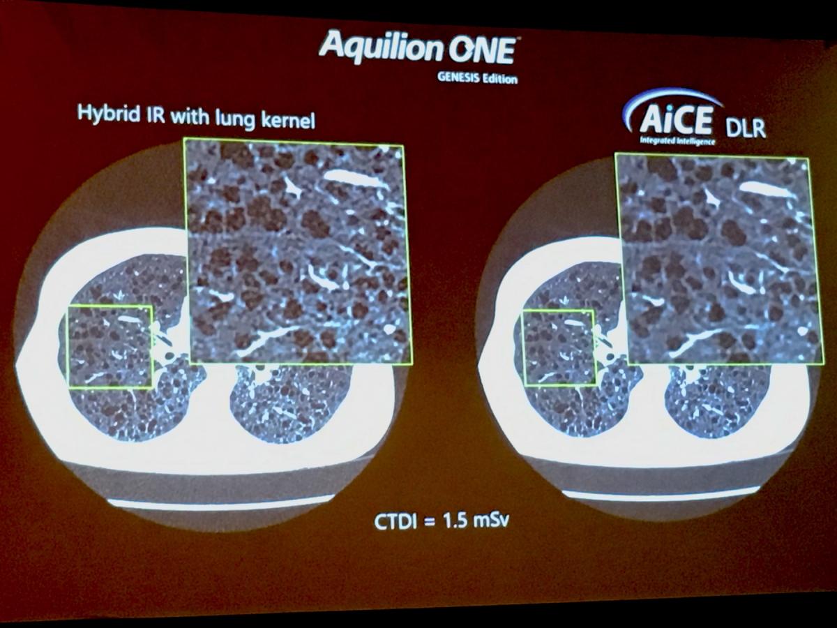
Vendors are developing new AI algorithms to reconstruct CT images better than conventional iterative or model-based reconstruction methods. Both Canon Medical Systems and GE Healthcare showed examples of their deep-learning CT image reconstruction software, both of which gained U.S. Food and Drug Administration (FDA) clearance in the spring of 2019.
 In deep learning, you feed the system the answers (in the case of CT, what ideal clinical images and resolution should look like) and the machine figures out what needs to be done with input image data to make it look like the ideal reference image, explained Jeannie Yu, M.D., FACC, FSCCT, director of cardiovascular imaging, Long Beach VA Health System. In this way, she said deep learning can be used to help in image resolution recovery that has better noise reduction and resolution than iterative reconstruction images.
Her center is using Canon Medical Systems’ AiCE deep learning reconstruction (DLR) software for the Precision CT system. The technology uses 10 convolutional neuro network (CNN) algorithms with different kernel sizes to reconstruct the images. Yu illustrated that this AI-driven software takes a lot of computing power, with the system operating using 71.2 teraflops. A teraflop is a unit of computing speed equal to one million floating-point operations per second. For comparison, Yu said IBM Watson AI software operates at 80 teraflops, a Playstation 4 game console at 1.8, Xbox One X gaming console at 6 and the iPhone X runs at 5 teraflops.
GE Healthcare also showed examples of its recently cleared Deep Learning Image Reconstruction (DLIR) software on the new Revolution Apex CT system. This next-generation image reconstruction option uses a dedicated deep neural network (DNN) to generate what GE calls TrueFidelity CT Images. Compared to current iterative reconstruction technology, TrueFidelity CT images can elevate every image to offer better image quality, image sharpness and noise texture, according to GE.
AI also is starting to be used to sort through large volumes of complex patient data in studies for cardiac CT. James Min, M.D., director of the Dalio Institute for Cardiac Imaging, Weill Cornell, said the CONFIRM Registry uses AI to examine 35 key data points to look for new ways to better risk-assess patients and find common markers for patients who had subsequent cardiac events and those who did not. Min said it takes a core CT lab about 6-8 hours to fully evaluate a CT exam for a trial, and that an imager reviews about 3.15 billion pixels of information per day looking through medical imaging studies. To sort through tens of thousands of CT exams to find commonalities that can be used for better risk assessments and to identify radiomic markers, he said AI will have to be used to deal with such vast numbers in a time effective manner.
2. CT Imaging for Transcatheter Mitral Valve Interventions
 For the past several years SCCT has hosted hands-on workshops for transcatheter aortic valve replacement (TAVR) CT image planning, but this year was the first where it held a dedicated workshop on transcatheter mitral valve replacement (TMVR). TMVR imaging requirements are much more involved than TAVR because of the mitral valve's complexity and greater anatomical variation. TMVR is expanding with several transcatheter valves and repair techniques entering clinical trials, and the FDA has now cleared indications of TAVR valves to be used in valve-in-valve (VIV) and valve-in-ring (VIR) procedures.
"Because this field is so new, there are still a lot of questions for which we still do not have at the answers, so we need to be transparent in what we do. I hope that over the next few years, we should have a white paper document on how we should expand the indications for this therapy as we build more confidence," explained Joao Cavalcante, M.D., FSCCT, director of structural heart CT and cardiac MRI, Minneapolis Heart Institute.
He was one of the workshop instructors and spoke in sessions about TMVR imaging. He shared insights in the VIDEO: Transcatheter Mitral Valve Replacement Planning.
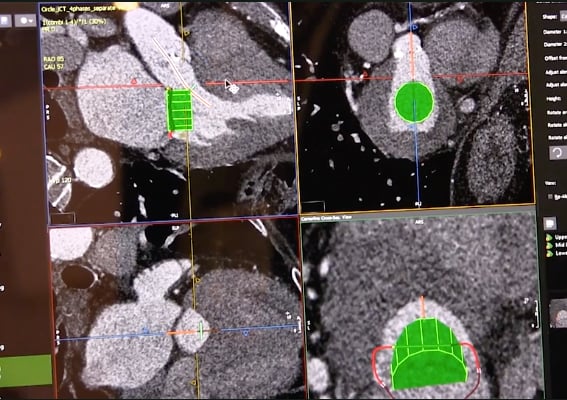
In addition to assessing anatomical access routes for catheters and device sizing, a key element of TMVR planning is calculating the area of the new left ventricular outflow tract (neo-LVOT) with the skirt of the TAVR valve hanging into the left ventricle. This has been a key point in all the sessions for TMVR, as it is a major predictor of patient outcomes and mortality. Vendors on the expo floor demonstrated several technologies to model the neo-LVOT from CT scans, including virtual placement of valves to determine proper landing zones and if additional work is needed to prevent blocking the LVOT, such as cutting the mitral valve leaflets using the LAMPOON procedure.
Another TMVR instructor, Dee Dee Wang, M.D., director of structural heart imaging, Henry Ford Hospital, Detroit, has done a lot of research on how to use CT imaging to prevent LVOT obstruction. She offers an overview in the VIDEO: The Importance of the Neo-LVOT in Transcatheter Mitral Valve Replacement.
Here is an example of TMVR software that was demonstrated on the expo floor at SCCT — VIDEO: Evaluation Tool for Neo-LVOT in Transcatheter Mitral Valve Procedures.
3. CT Calcium Scoring Now Part of Guidelines
 The American Heart Association (AHA) released updated cholesterol management guidelines late last fall that now encourage the use of CT calcium scoring risk assessment. It was one of the hot topics at the SCCT 2019 meeting and was discussed in dozens of sessions. The guidelines state calcium scans can be used to assess a patient's risk of having a coronary event. This risk assessment also can help patients and their doctors decide if statins should be prescribed. If a patient is on the fence or resistant to taking statins, thought leaders at SCCT say the calcium scan can be used to show the need (or no need) for statins. If a patient has a calcium score of zero, they do not have a risk for coronary events and there is no need for statins.
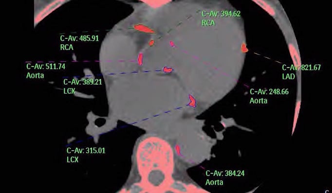
A low-dose CT calcium scoring scan can be performed to show the amount of calcified atherosclerosis in a patient's coronary vessels. Software automatically tallies the percentage of plaque in each vessel segment and compiles an Agatston score showing the risk of having a coronary event. Many hospitals now offer calcium scans at a low cost, between $50-$100, to screen patients and bring higher-risk patients into that cardiac care network.
 To find out more about this risk assessment DAIC spoke with Arthur Agatston, M.D., clinical professor of medicine, Florida International University, Herbert Wertheim College of Medicine, who is the namesake of the calcium scan Agatston scoring system. Watch the VIDEO: The History of CT Calcium Scoring.
Here is an interview with Matthew Budoff, M.D., professor of medicine, David Geffen School of Medicine, UCLA, at the AHA meeting last November when the guidelines were released — VIDEO: New Cholesterol Guidelines Support CT Calcium Scoring for Risk Assessment
4. Leveraging Social Media to Highlight Key Cardiology Articles
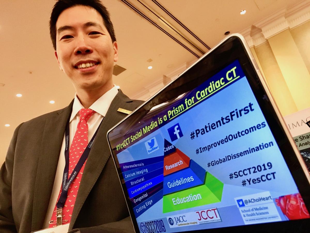 Several cardiology societies have started hosting regular sessions on how to leverage social media to more effectively share key news, published studies and new guidelines in the various cardiac subspecialties. SCCT started a big social media effort in 2018, including the creation of the hashtag #YesCCT for information on cardiac CT in appropriate indications. In a year since its introduction, #YesCCT has had more than 2,200 users, 11,000 tweets and more than 30 million impressions, explained Andrew Choi, M.D., FACC, FSCCT, co-director, cardiac CT and MRI, assistant professor of medicine and radiology, George Washington University, Division of Cardiology, Washington, D.C., who spoke in several sessions about use of social media in cardiology. He said use of specific hashtags enables social media to be used as a sort of vast journal club, with users across the world.
"To keep up with what is happening in the field, it is very difficult for any individual to read multiple journals a day, but what social media allows you to do is to access that content across multiple journals and make that connection and be able to go deeper on the articles that catch your interest," Choi said. "The role is really to be able to take what has been peer reviewed and published and is the best science and be able to translate and disseminate it to the cardiovascular and radiology communities. Social media allows them to easily engage in the information in bite-sized chunks so they can hopefully start to incorporate this information, know what is important in the field and take a deeper dive into those articles. This will help us propel the field forward."
Watch an interview with Choi in the VIDEO: Use of Social Media For Cardiology Education.
5. New Cardiac CT Scanner Technology
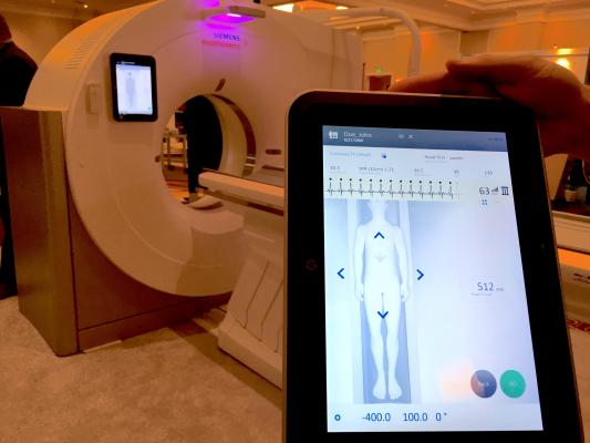
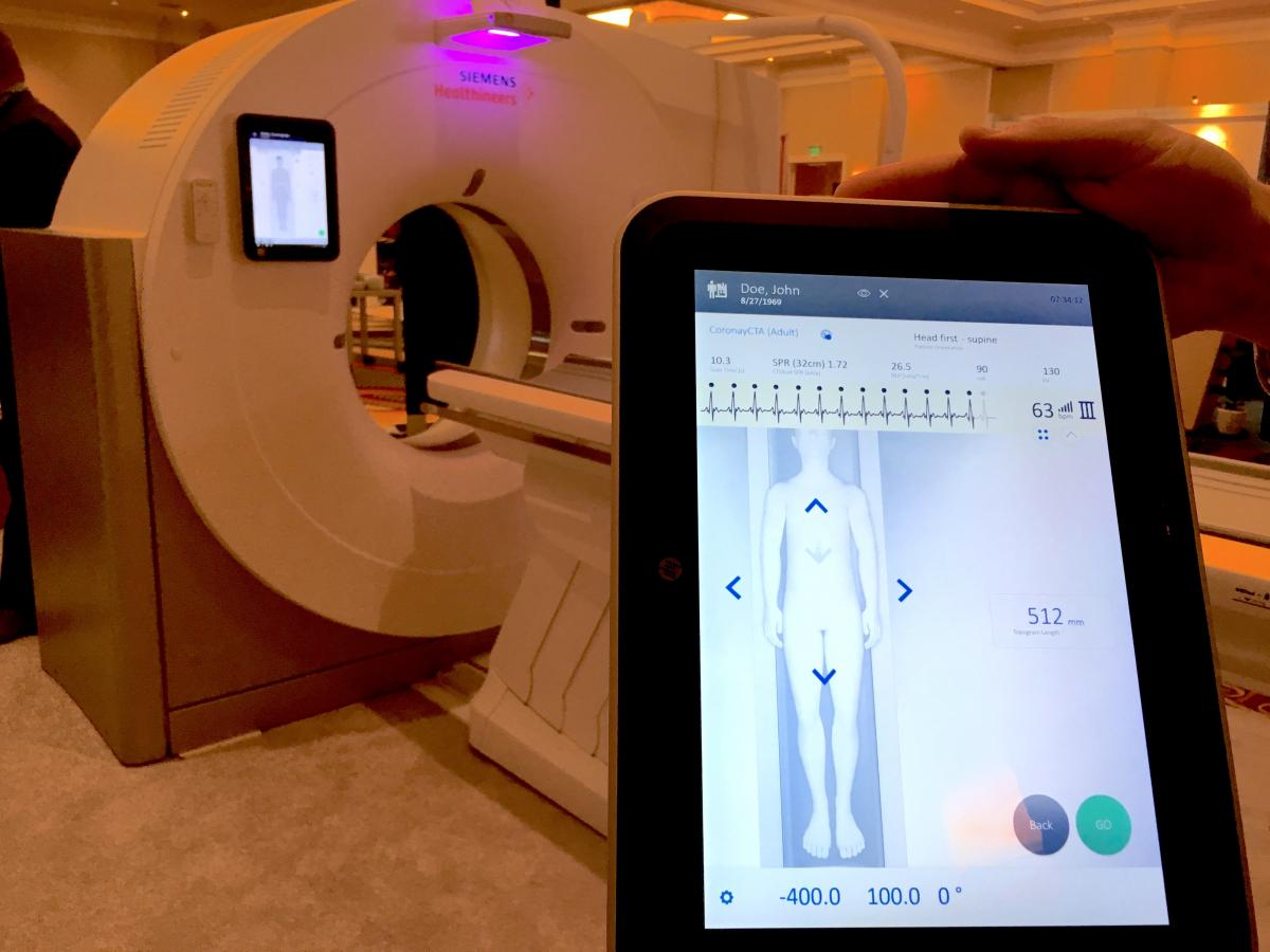 Siemens showed its Go.Top Cardiovascular Edition CT scanner. It has a compact CT gantry so it can fit in small rooms and is aimed at cardiology office-based imaging. It was released this past spring at the American College of Cardiology (ACC) meeting. The system has removable tablets on each side of the scanner which the tech can use to adjust the machine, review scout scans and trigger the scanner. The idea is to improve workflow and allow the tech to remain with the patient at the bedside longer, rather than tucked away in a remote control room using an intercom.
The entire system is built into the gantry, so there is no need for extra equipment in a closet, cabinet or server tower. It comes in a 128-slice configuration with 4 cm of anatomical coverage per rotation. It uses the Stellar detector and tin filtration to eliminate low-energy photons and help lower dose. It can be programmed to aid workflow by automatically removing bone, create cured planar reconstructions, lung computer-aided detection (CAD) and other post-processing features so more time can be spent on reading scans. The scanner also comes with a HeartFlow fractional flow reserve-computed tomography (FFR-CT) starter pack.
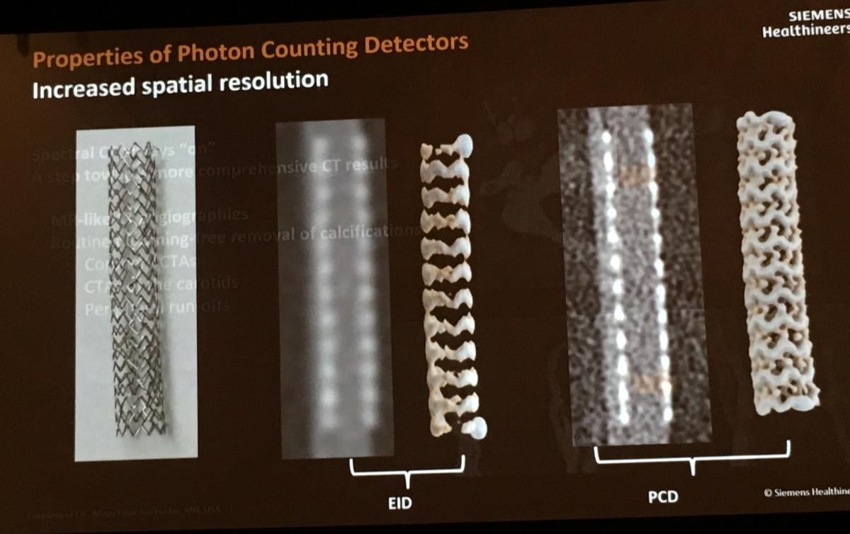 Siemens also showed examples of imaging using its photon counting detector, in development and being tested at three hospitals including Mayo Clinic. The vendor said this technology helps nearly eliminate electronic noise in images, improves tissue contrast, and allows for more MRI-like tissue characterization. The photon counting detector also can collect data using multiple energy thresholds, so it has intrinsic spectral imaging sensitivity.
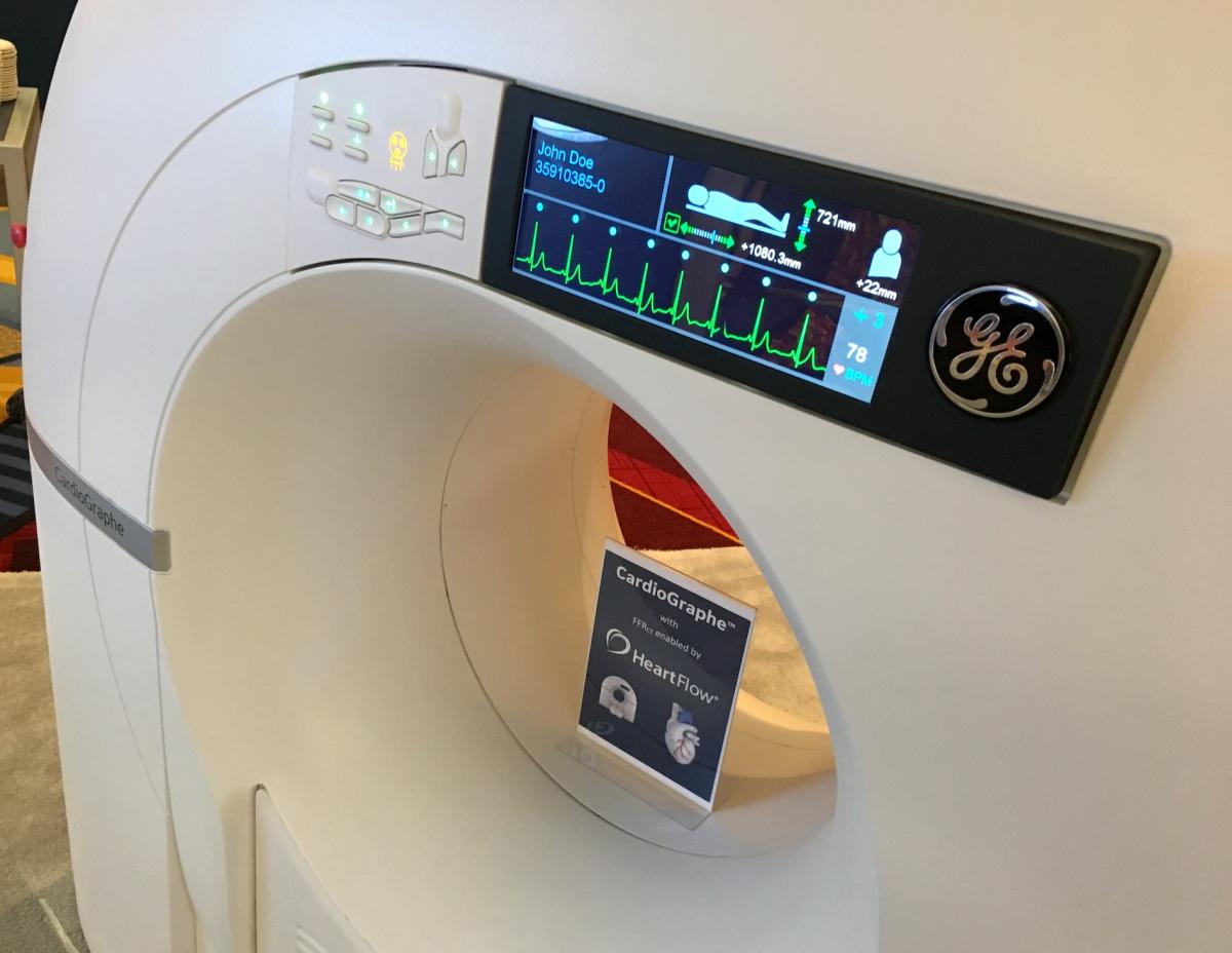 GE Healthcare showed its Cardiographe dedicated cardiac CT system at SCCT. It was designed specifically for cardiac imaging and so has a very compact footprint so it can be used in an office setting or small room. One of these systems was recently installed at St. Paul’s Hospital in Vancouver, Canada, where they have an extensive structural heart program. Read more about their use of this system.
6. Advances in Spectral Imaging
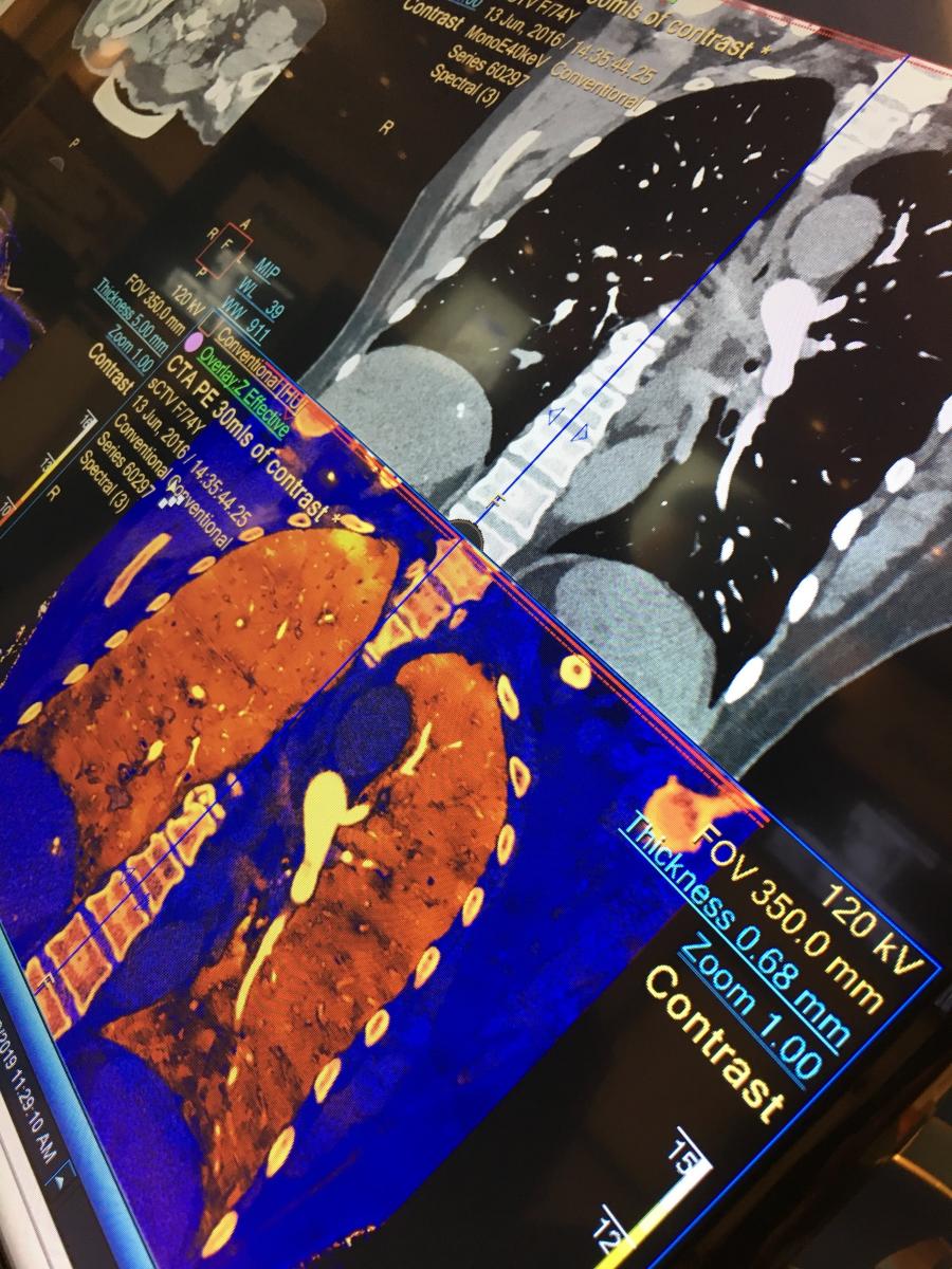 Spectral CT, also referred to as dual-source or dual-energy CT, has gained a lot of interest in the past few years because of its potential to offer additional information from CT images. This includes the ability to help definitively make a diagnosis in cases of small pulmonary embolisms, determining if shadowing inside a stent is artifact or restenosis, better delineation of aortic stent graft endoleaks, and to better visualize ischemia or infarcts using enhanced iodine mapping techniques based on different X-ray energies, explained outgoing SCCT President Suhny Abbara, M.D., FSCCT, in sessions. He is chief of cardiothoracic imaging and chair of the CT operations committee, University of Texas Southwestern Medical Center, Dallas, and is heavily involved in spectral imaging research.
Siemens, GE, Philips and Canon now all offer spectral imaging capabilities on some of their newest scanners. The vendors say there is increasing interest from imagers because of the extra information spectral CT offers.
See examples of spectral CT imaging and watch an interview with Abbara in the VIDEO: Applications of Spectral CT.
7. Special Sessions for EP and Structural Heart
SCCT has started partnering with other cardiology societies to host joint sessions aimed at specific subspecialties. It partnered with the Heart Rhythm Society (HRS) for a session on CT imaging for electrophysiology. Another session held jointly with Transcatheter Cardiovascular Therapeutics (TCT) was aimed at what interventional cardiologists need in CT imaging, especially in regards to structural heart planning for transcatheter valve implantations.
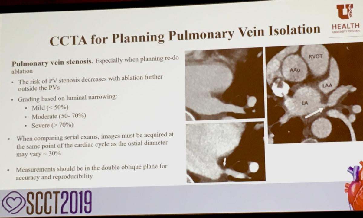 Mark Ibrahim, M.D., FACC, assistant professor of medicine and radiology, associate program director, advanced cardiac imaging fellowship, University of Utah, spoke on what radiologists and cardiologists need to know and what is needed from CT imaging prior to ablation procedures for atrial fibrillation (AF) and ventricular fibrillation (VF). Watch an interview with him in the VIDEO: What Electrophysiologists Need From CT Imaging Prior to AF and VT Ablations.
Pierre Qian, MBBS, an electrophysiology fellow at Brigham and Women’s Hospital in Boston, spoke about the work his center is doing in collaboration with radiation oncology to treat ventricular tachycardia (VT) in patients where their original catheter ablation did not work. They use the catheter electromapping from the previous catheter ablation and a CT scan to create a treatment plan for the radiotherapy system. Watch the VIDEO: Use of Radiotherapy to Noninvasively Ablate Ventricular Tachycardia.
Other SCCT 2019 Videos:
VIDEO: 10 Tips to Improve Cardiac CT Imaging — Interview with Quynh Truong, M.D., director of cardiac CT, NewYork Presbyterian Hospital
VIDEO: Walk Around of a Siemens Go.Top Dedicated Cardiac Scanner
VIDEO: Walk Around of a GE Cardiographe Dedicated Cardiac CT Scanner
Image Gallery From the Society of Cardiovascular Computed Tomography 2019 Meeting
VIDEO: Example of Photo-realistic CT Imaging of a CoreValve
VIDEO: Example of an Automated CT Cardiac Calcium Scoring Exam
VIDEO: Example of In-Stent restenosis Visualized Using Spectral CT
VIDEO: Realistic Cinematic Reconstruction of a Heart CT Scan
Find more news and video from SCCT.


 February 06, 2026