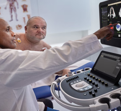
The Philips Epiq cardiac ultrasound system includes automated measurements and anatomical identification based on an artificial intelligence algorithm.
Cardiac ultrasound technology is undergoing an automation technology transformation similar to that seen with computed tomography (CT) advanced visualization software over the past decade. For years, echocardiography software tools like strain and 3-D assessment have existed to help in cardiac quantification, but the technology required numerous steps, was time-consuming and often had to be done on a separate workstation. Additionally, there was wide variability in how the exams were scanned and how measurements were taken based on user experience. Today, the latest generation echo systems are introducing a high level of intuitive workflow and automation, including artificial intelligence, to increase reproducibility and significantly reduce the time it takes for quantification.
As vendors have adopted “keep it simple” principles, technologies that were limited to luminary academic centers are now becoming easier to use, faster and more readily available in mainstream cardiac ultrasound systems.
“I was impressed seeing vendors able to put these technologies on the carts, and allowing you to be able to use it, on the cart while sitting with a patient. It’s a big step in the right direction to be able to use these new tools, advanced measurements and calculations we need,” said Sabrina Newell, MS, RCS, clinical analyst with market research company MD Buyline. “You can monitor the patients better and get better quality exams and better reproducibility.” She said the key to new technology adoption is for the sonographers to be comfortable in using it, and she has seen a lot of improvements in this area over the past year.
Expanding Adoption and Automation of 3-D Cardiac Ultrasound
There was initially slow uptake of 3-D cardiac ultrasound systems, partly because of its expense and complexity over traditional 2-D echo. However, that is slowly changing with advancements in automation to make 3-D assessments faster, easier and more accurate.
“We are definitely seeing more acceptance of 3-D,” said MD Buyline clinical analyst Jon Brubaker, MBA, RCVT. “It gets down to an ease-of-use workflow issue. The images are obviously extremely valuable.”
Unlike 2-D, where sonographers had to take several 2-D views of the heart to make measurements on each plane, one major advantage of 3-D is that it acquires full-volume datasets of the heart. This is similar to a CT or magnetic resonance imaging (MRI) where there is no inter-operator variability in the image acquisition, because there is no technique or experience required to get an exact plane and angle for a 2-D image. Since a volume dataset includes an image of the entire heart, it allows the operator, or smart software, to select any view required from one scan. With the use of advanced visualization software, 3-D datasets can be reformatted on any plane or angle. Additionally, some systems are sophisticated enough to automatically identify the anatomical structures in the image and extract optimal views. These systems also can automatically perform measurements and full-volume qualifications of the heart, rather than just basing numbers on a few select views of the heart. These features can greatly reduce issues with reproducibility.
A couple years ago, Philips began integration of artificial intelligence that automatically takes the image volume data and recreates the optimal diagnostic views. The Anatomically Intelligent Ultrasound (AIUS) suite technology was introduced on Philips' Epiq 7 cardiovascular ultrasound system at the American Society of Echocardiography (ASE) 2015 annual meeting. AIUS also uses the 3-D dataset to automatically compute quantification measurements to help clinicians quickly and easily assess disease states and determine treatment with highly reproducible results.
There is an ongoing effort to find ways to increase reproducibility and reduce variability in cardiac measurements. This includes new echo guidelines that provided updated normal ranges for chamber quantification and more detail on the standardization of measurement parameters. Echo experts advocate that patients should be able to see different physicians or be imaged by various sonographers and on different imaging systems without significant variability in their cardiac measurements.
Many vendors now offer on-cart advanced imaging technology and intuitive quantification software that can automatically assess ejection fraction and simplify myocardial strain analysis. By using the advanced software applications available and following a standardized measurement protocol, Brubaker said it is fair to presume these factors can reduce variability and increase the reproducibility of the ultrasound exam.
Increased Use of Strain Imaging
The use of strain imaging has grown in recent years as the technology has improved. It can provide objective parameters to the otherwise subjective determination of global and regional wall motion. Daily use of this tool is now becoming a part of many programs for assessing several disease states, including coronary heart disease, valvular heart disease, heart failure and cardiotoxicity tracking.
“Strain provides a great tracking mechanism that is really gaining acceptance,” Brubaker said. “Strain has been around for many years, but it has improved and there is now standardization among the vendors, and it is becoming easier to use.”
One of the biggest improvements is real-time strain imaging. Prior to current-generation echo systems, strain had to be done in post-processing off-cart, which could be very time-consuming. This did not fit well within a sonographer’s workflow, said Newell. She added that reproducibility of the results also varied by operator experience and where they took the measurements.
However, Newell said the latest systems make it easier to use, making the technology less complex and intimidating. One system showcased at ASE 2016 included a simple five-step process to automate the measurement of strain and intramural thickening.
Watch the VIDEO "CRT Optimization Using Echo," an interview with Leyla Elif Sade, M.D., MESC, professor of cardiology at Başkent University, Ankara, Turkey, on the use of echo for CRT optimization at the 2017 ASE annual meeting.
Watch the VIDEO "Assessing Cardiotoxicity Response With Cardio-Oncology Echo Imaging," an interview with Federico Asch, M.D., FACC, FASE, associate director of the echocardiography core lab at Medstar Health Research Institute and assistant professor of medicine (cardiology) at Georgetown University.
Evolution of Smaller, Compact Systems
The advanced features of premium cardiac ultrasound systems are trickling down to newer intermediate-level and compact, portable systems. These new software capabilities on portable systems include speckle tissue tracking for myocardial strain, auto quantification tools, real-time 3-D/4-D functionality, and the ability to perform live transesophageal echo (TEE) at the bedside or in the cath lab.
As 3-D ultrasound sees increased use in the cath lab and hybrid OR, some vendors are introducing new, compact systems. A good example of this is GE Healthcare’s Vivid iq, introduced in the fall of 2016. It is a small, mobile system with 3-D and has 3-D/4-D TEE capabilities for interventional procedures. It weighs 4.5 kg and matches the high image quality functionality of larger GE dedicated cardiovascular echo systems. It has a one-hour battery life and an easily transportable cart. It was designed for easy use and offers intuitive touchscreen functions to reduce the number of keystrokes needed.
Differentiating Features on Echo Systems
Here are several considerations clinical buyers should ask when assessing new systems:
• Does the system offer good ergonomics?
• Does the system offer workflow improvements that will reduce scan times or image quantification? This may include a reduction in steps to perform a function, reducing the number of clicks, or one-button automation features. Touch-screens are often used to help speed and simplify workflow.
• Is the system intuitive to use?
• How fast is the system’s image processing and how much image data is being processed?
• What technologies are used to improve image quality (i.e. how much data is being returned per slice; does the system use new beam-former technology to increase penetration, spatial resolution, noise reduction or contrast resolution?)
• What software features are offered?
• Is the system suited for contrast imaging?
• Can it perform fetal or pediatric imaging?
• Can it perform stress echo?
• Is the system enabled for TEE?
• If using TEE for interventional procedure guidance, can the images be fused with live fluoro?
Cardiac Ultrasound Systems Comparison Charts
DAIC has created an apples-to-apples comparison chart of technical specifications of both echocardiography and point-of-care ultrasound systems used for cardiac evaluation. The chart requires a login, which is free and only takes a minute to sign up. It can be accessed at www.dicardiology.com/content/cardiovascular-ultrasound
Video Tours of Vendors' Systems:
Watch the VIDEO “GE Healthcare Cardiovascular Ultrasound for Interventions.”
Watch the VIDEO “Siemens True Volume TEE, The Next Step in 3-D Imaging.”
Watch the VIDEO “Toshiba, See the Unseen for Early Detection in Cardiac Echo.”
Additional Echocardiography Technology Resources:
Read the article “Top Trends in Cardiac Ultrasound.”
Watch the VIDEO “Trends and Advances in Echocardiography at ASE 2016.”
VIDEO: Advances in cardiac ultrasound at the American College of Cardiology (ACC) 2016 meeting
Find links to news in “Key Technology Highlights in Echocardiography at ASE 2016.”
Watch video “What is Required for Interventional Echo - Discussion With Dr. Rebecca Hahn”


 June 15, 2026