Cases of acute cardiovascular disease and cardiac complications caused by COVID-19 require cardiovascular imaging require cardiac imaging to continue under the pandemic. All the key cardiovascular imaging societies in March ands April issued COVID-19 guidelines and lists of considerations for how to continue imaging operations safely under the threat of viral contamination during the duration of containment efforts.
Cardiovascular disease is the comorbidity that has carries the highest death rate in COVID-19 patients, at about 10.5 percent. For this reason, risk of cardiovascular complications in the setting of COVID-19, including pre-existing cardiac disease, acute cardiac injury and drug-related myocardial damage, will require the use of cardiac imaging, which will place cardiovascular imagers in the front lines of COVID-19 patient care.
All of the guidelines share similar key points for best practices to maintain safety and minimize contamination, triage of who should be imaged and who should be postpone to a later date, maintaining social distancing, hand washing, and cleaning of equipment. However, these guidelines vary slightly based on the specifics of each modality.
Across all imaging and procedures, all elective procedures have been postponed and how to triage patients is a key element of all the various guidelines.
"The tests should be done, very simply, if it changes the care of the patient. If it doesn't change the care of the patient, and it can be postponed, it should be postponed," explained Stephen Bloom, M.D., FASNC, director of noninvasive cardiology (cardiac CT, nuclear cardiology and echocardiography) at Midwest Heart and Vascular Associates. He is also a member of the American Society of Nuclear Cardiology (ASNC) Board of Directors. "I would say 80 percent of our cardiac imaging exams have stopped, it has been very dramatic.”
Right now, Bloom said it is difficult to test everybody and there is a shortage of masks, gowns and other personal protective equipment (PPE), and the imaging equipment needs to be sanitized each time it is used, so it just is not possible to image all the patients who need imaging right now. Hospitals also are trying to limit the number of healthy people people coming into hospitals for routine visits and tests to reduce their potential exposure to the virus and help containment efforts.
Common themes in all the recommendations from all the societies include:
• A screening checklist.
• Examples of how to triage patients for cardiology studies.
• Employ social distancing.
• Rescheduling non-urgent visits.
• Rescheduling elective surgeries and procedures.
• Using separate spaces for patients with known or suspected COVID-19 to prevent spread.
• Ensuring supplies are available.
• Promoting use of telehealth.
• Screen staff, patients and visitors before they enter the department.
• Minimize non-essential visitors into the department.
• Record symptoms at the start of the shift.
• Record temperature daily as per local policies and standards.
• Use personal protective equipment (PPE) for healthcare personnel.
• If available, use PPE for patients (due to concern of asymptomatic transmission of COVID-19).
• Maintain strict hand hygiene.
• Maintain 6 feet distance in all patient/staff interactions when possible.
• Minimize crowding in workplace.
• Work remotely whenever feasible.
• Use of virtual conference tools for meetings and educational conferences.
• Rotating staff schedules for on-site and off-site work.
• Training in local infection control recommendation.
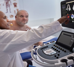
Recommendations for Echocardiography During COVID-19
The American Society of Echocardiography (ASE) statement said carefully consideration should be given to who should be imaged, where to image and how to image, since these three questions have the potential to reduce the risks of viral transmission.[1]
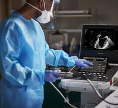
Since cardiac ultrasound require close contact with patients, the ASE statement urges judicious use of personal protective equipment (PPE), following best practices, pre-planning, exams and thoroughly sterilizing equipment after each exam and use of probe covers.
The use of transesophageal echo (TEE) should carries a heightened risk of spread of the COVID-19, because it can promote the aerosolization of a large amount of virus due to coughing or gagging that may result during these exams.
The ASE statement calls for cautious consideration of the benefit of a TEE exam should be weighed against the risk of exposure of healthcare personnel to aerosolized virus in a patient with suspected or confirmed COVID-19. These exams also require use of higher level PPE. ASE said TEEs should be postponed or canceled if an alternative imaging modality can be used, including off-axis transthoracic echo (TTE) views, ultrasound enhancing agent with TTE, or use of contrast enhanced computed tomography (CT) and magnetic resonance imaging (MRI). CT and MRI have emerged as an alternative to TEE for exclusion of left atrial appendage (LAA) thrombus prior to cardioversion, for example, the authors state.
The use of alternative tests to avoid an aerosolizing procedure should be balanced against the risk of transporting a patient through the hospital to the CT or MRI scanner, the need to disinfect the CT or MRI room, and iodinated contrast and radiation for CT, or long scan times for MRI. The authors stated some institutions have a dedicated CT scanner reserved for patients with COVID-19.
In addition to limiting the number of echocardiography practitioners involved in scanning, consideration should be given to limiting the exposure of staff who may be particularly susceptible to severe complications of COVID-19. Staff who are more than 60 years old, have chronic conditions, are immunocompromised or who are pregnant may wish to avoid contact with patients suspected or confirmed to have COVID-19, depending on local procedures, the statement reads.
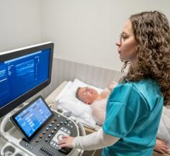
Numerous hospitals have found use of very small point-of-care ultrasound (POCUS) systems based on mobile phones or iPads combined with a transducer and an app allows for quick answers to clinical questions. These systems are small enough to be put into protective sheath and are much easier to clean than a larger ultrasound unit. Dedicated POCUS devices are often being used for COVID-19 patients and left in the ICU to limit cross contamination.
Best Practices for Nuclear Cardiology Laboratories During COVID-19
A guidance document for nuclear imaging labs was created in partnership between the American Society of Nuclear Cardiology (ASNC) and the Society for Nuclear Medicine and Molecular Imaging (SNMMI). The "Guidance and Best Practices for Nuclear Cardiology Laboratories During the Coronavirus Disease 2019 (COVID-19) Pandemic: An Information Statement from ASNC and SNMMI," offer specific recommendations for adapting nuclear cardiology practices at each step in a patient’s journey through the lab — for inpatients, outpatients and emergency department patients.[2]
Specific nuclear imaging recommendations include:
• Use of rest only studies if possible to reduce exam time and avoid increased viral shedding from stress exam respiration.
• Use of half-time SPECT to speed exam times.
• Use of PET if available to speed exam times.
The document offers much more detail on these topics and how nuclear cardiology departments can adapt to the new situation, while still accommodating patients.
COVID-19 Considerations in Computed Tomography
Computed tomography (CT) emerged as a front-line imaging modality in China early in the pandemic. CT is able to clearly show pneumonia and can help confirm COVID-19 cases, sometimes before a positive reverse-transcription polymerase chain reaction (RT-PCR) test, or gene sequencing, for respiratory or blood specimens.[3]
"With the superior images and new technology now allowing for physiologic assessments, we have really seen CT emerge as a front-line test for the evaluation of chest pain, and now even more so in the era of COVD," explained Geoffrey Rose, M.D., president of Sanger Heart and Vascular Institute with Atrium Health, in Charlotte, N.C. "We have found the ability of CT to be able to get patients in and out the door has enabled us to image patients faster and with less physician contact is well suited to the COVID environment."
For this reason, the Society for Cardiovascular Computed Tomography (SCCT) explained in its COVID-19 guidelines that incidental lung findings in CT angiography (CTA) are important and should be reported immediately.[4]
SCCT said cardiac CT imaging will likely show incidental pulmonary findings in patients at risk of COVID-19 exposure. COVID-19 is a viral pneumonia, with a spectrum of findings ranging from normal lungs to acute respiratory distress syndrome. SCCT said typical chest CT findings in known cases are described in the reference article “Radiology Department Preparedness for COVID-19: Radiology Scientific Expert Panel," published in Radiology,[5] and on the British Society of Thoracic Imaging: COVID-19 Resources webpage.[6]
If typical or atypical pulmonary findings are encountered, SCCT suggests consultation with a radiologist with thoracic expertise, appropriate documentation and timely communication of these findings, especially in cases not known or suspected to have COVID-19.
The SCCT document lists precautions to follow:
• Encourage sick employees to stay home. Personnel who develop respiratory symptoms (e.g., cough, shortness of breath) or unexplained fever should be instructed not to report to work.
• Ensure that your sick leave policies are flexible and consistent with public health guidance and that employees are aware of these policies. Make contingency plans for increased absenteeism
• Screen patients and visitors for symptoms of acute respiratory illness (e.g., fever, cough, difficulty breathing) or gastrointestinal symptoms and coronavirus exposure in the last 2 weeks before entering one’s healthcare facility.
• Consider standard droplet precautions for patients and healthcare personnel as per institutional infection control protocols.
• Increase scheduling intervals or appointment times to allow adequate time to clean equipment as needed.
• Leverage telemedicine technologies and isolated workstations to allow for reading and interpretation, that allow for social distancing to limit staff exposure, when possible.
• Assign a team member to monitor and incorporate regular updates from the CDC and appropriate regional jurisdictions.
• CCT may be preferred to transesophageal echocardiography (TEE) in order to rule-out LAA and intracardiac thrombus prior to cardioversion in order to reduce coughing and aerosolization related to TEE.
• The ability of CCT to decisively exclude coronary disease or high-risk anatomy may prevent the need for inpatient admissions and resource use.
• Consider that elderly patients, those with co-morbidities, and those who may be immunosuppressed are at greater risk of morbidity / mortality from COVID-19, and the benefit and risk of cardiac CT should be evaluated on a case by case basis.
• In patients under investigation (PUI) and with confirmed COVID-19, the benefit of CCT in most clinical scenarios will likely be lower than the risk of exposure and infection to healthcare
CT imaging during COVID-19 of suspected or positive patients requires appropriate environmental cleaning and decontamination of CT rooms after each patient. SCCT said this requires thorough cleaning of the surfaces by a staff member with appropriate PPE as per CDC and local institutional guidelines for airborne viral diseases.
SCCT's document outlines specific cardiovascular CT protocol triage considerations for common indications during COVID-19. To explain elective indications can be delayed more than 8 weeks, semi-urgent cases should rescanned within 4-8 weeks, and urgent indications should be scanned in less than 2-4 weeks.
Related Imaging Precautions During COVID-19 Content:
Best Practices for Nuclear Cardiology Laboratories During the Coronavirus (COVID-19) Pandemic
ASE Guidelines for the Protection of Echocardiography Providers During the COVID-19 Outbreak
VIDEO: Best Practices for Nuclear Cardiology During the COVID-19 Pandemic — Interview with Hicham Skali, M.D.
VIDEO: Cancelling Non-essential Cardiac Procedures During the COVID-19 Outbreak — Interview with Ehtisham Mahmud, M.D.
VIDEO: 9 Cardiologists Share COVID-19 Takeaways From Across the U.S.
VIDEO: Telemedicine in Cardiology and Medical Imaging During COVID-19 — Interview with Regina Druz, M.D., FASNC, a member of the American Society of Nuclear Cardiology (ASNC) Board of Directors, chairwomen of the American College of Cardiology (ACC) Healthcare Innovation Section
VIDEO: Use of Teleradiology During the COVID-19 Pandemic — an interview with John Kim, M.D., chairman, Department of Radiology, THR Presbyterian Plano, Texas
VIDEO: Imaging COVID-19 With Point-of-Care Ultrasound (POCUS) — Interview with emergency physician Mike Stone, M.D.,
VIDEO: How China Leveraged Health IT to Combat COVID-19 — Interview with Jilan Liu, M.D., CEO for the HIMSS Greater China
Study Looks at CT Findings of COVID-19 Through Recovery
Experts Stress Radiology Preparedness for COVID-19
ACR Recommendations for the Use of Chest Radiography and CT for Suspected COVID-19 Cases
VIDEO: What Cardiologists Need to Know about COVID-19 — Interview with Thomas Maddox, M.D.
The Cardiac Implications of Novel Coronavirus
References:

 July 02, 2026