Over the past several years, the focus on new technology at the annual Radiological Society of North America (RSNA) scientific meeting in Chicago has moved away from new devices to new software. RSNA 2010, held Nov. 28 through Dec. 2, emphasized this trend. The main technical advances displayed expand and simplify computer access to images, offer new advanced visualization tools, improve image quality using new processing algorithms and help lower radiation dose.
The newest trend at this year’s show was the integration of picture archiving and communication systems (PACS) and advanced visualization software into the iPad. It seemed every major vendor was showing new compatibility with iPad personal computing devices, allowing nearly instantaneous, wireless access to any patient’s images and reports from anywhere. This introduces a new concept of what constitutes a workstation and unchains the radiologist or cardiologist from his or her desktop PACS. Many vendors also believe being able to call up images anywhere will enhance collaboration between physicians in different hospital departments, referring physicians and patients.
Two key advances were also seen with regard to imaging hardware. The biggest was the unveiling of two positron emission tomography/magnetic resonance imaging (PET/MRI) systems by Philips and Siemens. These systems have the potential to greatly reduce radiation dose by eliminating CT scans in hybrid imaging.
Cassette-based computed radiology (CR) also seems to be on the way out with the introduction of affordable digital radiography (DR) retrofit systems. In addition, second-generation wireless DR detectors are being introduced to untether and untangle X-ray rooms.
The Coolest Finds on the Show Floor
Here is my list of the most interesting technologies I found on the RSNA technical exhibit floor. I based the coolness factor on being unique and offering new insights into how to improve workflow, clinician education and new ways to view images.
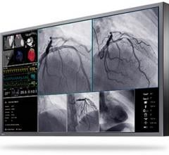
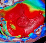
3-D Hearts in Motion
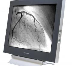
Advanced visualization can make pretty pictures of 3-D anatomy, but in the case of the heart, it has so far failed to show functionality and natural movement during the cardiac cycle. PACS users can flip through sets of stacked images from a CT or MRI data set to make a sort of primitive cartoon, but it lacks the smooth flow of real anatomical motion.
Using supercomputing algorithms previously used to forecast weather, Ziosoft developed the PhyZiodynamic software to take a CT or MRI data set and put it into motion. The software has an option to show a color-coded tissue strain map. At ACC last March, the software took about eight hours to process a cardiac cycle dataset, which limited its practical application. However, the company has cut processing down to about 45 minutes and is working to reduce it further.
At its RSNA booth, Ziosoft offered a full 3-D rendering of a heart in motion and invited attendees to rotate and virtually dissect it. The beating heart could be sliced through while in motion, offering views of the myocardium and valves in motion as only previously seen by surgeons. The views also showed the movement of the valve leaflets and the chordae tendineae.
The application of this technology holds promise in surgical planning, transcatheter valve replacements and septal shunt occlusion. It may also allow a new breed of hybrid imaging in the cath lab, where live angiography could be synchronized with a 3-D cardiac reconstruction in motion, rather than a frozen image.
The company hopes to have a commercialized version of the software in 2011.
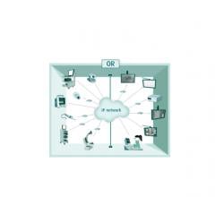
An iPad that Controls PACS Workstations
While many vendors at RSNA showed integration of their PACS and advanced visualization functionality with the iPad, McKesson’s engineers took the concept a step further. The company showed works-in-progress software that enables an iPad user to take the device to any workstation in a hospital and wirelessly connect with it. The iPad then becomes the controller for what is viewed on the larger, diagnostic-quality display screen. It allows the touchscreen and figure movement workflow that make the iPad and iPhone-type devices so popular. The advantage of the system is it allows quick access and viewing on diagnostic-quality screens, which will please the FDA, since iPad screens are not considered high enough quality from which to make a diagnosis.
When radiologists first switched from film to PACS-based image review systems, many complained the computer forced them to stay at a desk in one location all day, rather than taking an image with them to show other physicians involved in a patient case. Many say the conversion to PACS reduced the level of face-to-face collaboration. This new method of carrying images around may return some of this lost face-to-face time with colleagues.
McKesson said RSNA attendees really liked the concept of the device interface and the company plans to move forward with commercializing the technology in 2011.
PET/MRI Makes its Debut
Both Siemens and Philips showed their works-in-progress PET/MRI systems. While the modalities are the same, they differ in their designs.
The major obstacle to developing this hybrid imaging system has been that photo-multipliers used in PET imaging do not operate in intense magnetic fields. Siemens engineers figured out how to shield the system to integrate the PET detectors inside the MRI machine. The result is a machine a little longer than a standard MRI system.
Philips solved this issue by separating the two imaging systems on either side of a room, separated by a rotating platform with the patient table. The system first loads a patient into the MRI scanner, then pulls him or her out, rotates 180 degrees and loads him or her into the PET scanner. The computer system then aligns the two images.
In terms of size, the Siemens system can be installed in a standard radiology room, whereas the Philips system may require the area of about two rooms. The Siemens system obtains both the PET and MRI images simultaneously, whereas the Philips system does not.
Siemens is hoping for European CE mark approval in 2011. The Philips system is currently pending FDA 510(k) approval, which the company hopes will come sometime in 2011.
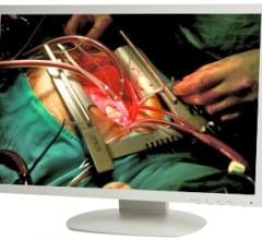
The ‘CSI’ Workstation
Sectra introduced its new Visualization Table, a 46-inch medical multi-touch display, where multiple users can interact collaboratively with real-size, 3-D images generated by CT and MRI scanners. The table-sized display and its use of hand and finger motions to manipulate images, looks just like a prop of the hit TV shows “CSI” or “Bones.” The company envisions it being used for quick review by several physicians in multi-trauma cases. It may also have applications for medical education, clinical conferences and virtual autopsies.
New Generation of Educational Aids
Two educational aids caught my eye at RSNA. Most clinicians are familiar with the Body Worlds exhibits of cadavers preserved using a proprietary preservation method that turns tissue into a polymer. Gubener Plastinate displayed preserved 1-5 mm cross-sectional body slices of various parts of the human anatomy. The specimens offer a true, detailed histological view of real anatomy that is usually only seen on CT or MR slices, but with much more detail.
Another interesting educational aid from Anatomage was The Table, made of two diagnostic-quality displays preloaded with a life-sized, full-body data set scan of a patient, finished off with a photo overlay skin. The data set can be sliced to show the internal anatomy and bones. The virtual body can also be rotated or “dissected” on any axis with hand and finger movements. The system is designed to better educate clinicians about anatomy in a more interactive way, instead of relying on traditional 2-D textbook illustrations.
For more information: www.rsnafastpass.com/rsna2010


 January 25, 2018