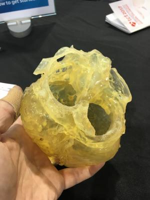
The use of 3-D printed hearts from patients' pre-TAVR planning CT scans have improved outcomes of procedures at the University of Minnesota. Clearly identifying where calcium is located on the valves prior to TAVR device implantation has helped reduce the incidence of paravalvular leak.
May 14, 2018 – A new study examines the effectiveness of 3-D printing technology and computer modeling to predict paravalvular leak (PVL) in patients undergoing transcatheter aortic valve replacement (TAVR). A common risk of TAVR is an ill-fitting valve which can lead to PVL. To address this risk, the study used 3-D printing technology to help confirm and detect the location of the leak. The retrospective study was presented today at the Society for Cardiovascular Angiography and Interventions (SCAI) Scientific Sessions.
More than 5 million Americans are diagnosed with heart valve disease each year.[1] TAVR is a procedure used for intermediate, high-risk, and inoperable patients with severe narrowing of the aortic valve where a prosthetic valve is implanted and the damaged valve is replaced. Patients who undergo TAVR, which is a less invasive procedure to replace the heart’s aortic valve, can experience paravalvular leak around the new valve which can lead to higher mortality rates. Therefore, clinicians are exploring ways to find and prevent these leaks from happening. 3D printing has become more popular within the medical space as it has been discovered to be a vital tool to prevent, fix and foresee procedural errors.
In the study, six patients undergoing TAVR for severe, calcific aortic stenosis and at risk for paravalvular leak had pre-procedure computed tomography (CT) images analyzed and segmented for printing of 3D models. The CT scans allowed researchers to see a 360-degree view of the location of the calcium build up while the 3D models allowed researchers to further evaluate the ill-fitting valves. The 3D aortic root models were then implanted with the valve to determine if the size was correct, ultimately revealing where the calcium composites would be. The 3D models were scanned, evaluated for final analysis and then compared to in-vivo implanted TAVR echocardiograms.
Every leak seen on the 3D models were confirmed on the CT digital scans. The 3D models allowed researchers to use prototypes to personalize valve placement, size and location to stop leaks and lower calcium build up.
“We are very encouraged to see such positive outcomes for the feasibility of 3D printing in patients with heart valve disease. These patients are at a high risk of developing a leak after TAVR, and anything we can do to identify and prevent these leaks from happening is certainly helpful,” said lead author Sergey Gurevich, M.D., and cardiovascular fellow at the University of Minnesota in Minneapolis, Minn. “Like any other new technology, as 3D printing evolves, we hope to see an increase in accessibility and opportunity for the use of this technology to help improve patient care.”
The authors call for a functional study to help determine the exact size of the leak. The authors of this study are working with computational fluid dynamics to optimize calculations.
Watch the VIDEO "Applications in Cardiology for 3-D Printing and Computer Aided Design" — interview with Dee Dee Wang, M.D., Director, Structural Heart Imaging at Henry Ford Hospital, Detroit.
Complete listing of SCAI 2018 late-breaking trials with links to articles.
Reference:

 June 18, 2026


