ScImage’s PICOM365 Enterprise PACS enables cath, echo, vascular, ECG, stress and Holter storage and reporting.
Call nights are by far the worst part of being a clinical cardiologist. I love what I do during the day, but on weekends where I am taking clinical calls for my six-man (one woman) cardiology group, as well as covering weekend echo and transesophageal echo (TEE) for my hospital, the stress can be high.
With the advent of efficient cardiac PACS, however, many of the stressful elements have gone away. In particular, the ability to remotely read and report on cardiac imaging studies has revolutionized my workflow. Now the need to get in my car and drive to the hospital to view studies has been eliminated. Leaving the hospital is no longer dependent on all work being completed.
On a recent Friday evening on a call weekend, my Eternal Fiancée insisted that we go to Oceano for happy hour. Oceano is a Clayton, Mo., seafood restaurant that arguably has the finest happy hour in the western world. In the dark era preceding electronic health records (EHR) and online PACS, this would have been out of the question.
As we were walking out the door, endeavoring to reach Oceano before happy hour ended, the sonographer on call texted me that he was going in to perform a STAT echo on an ICU patient to rule out pericardial effusion. In the dark era, this would have been devastating. The Oceano visit would have been canceled and I would have had to drive in to the hospital.
When I started at St. Luke’s 10 years ago, our cardiac cath lab stored their cath films on ScImage’s PICOM365 Enterprise PACS, and we have subsequently added echo, vascular, ECG, stress and Holter storage and reporting.
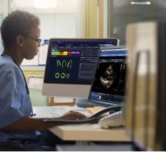
I can easily access any patient study, no matter the discipline. The first time I read an on-call STAT echo using PICOM365 Cloud technology on my iPad in a restaurant, my mind was blown. It was as if a heavy weight had been lifted from my on-call duties.
Now, 10 years later, I’m eating Oceano’s delicious blackened big eye tuna (rolls of ahi tuna filled with radish, shiitake mushrooms, pickled ginger and scallions with spicy mustard sauce) when the sonographer texts me that the STAT echo is ready and that he is concerned about a pericardial effusion. I had brought my MacBook Pro with me (yes, hospitals have finally figured out that doctors often use Macs), so I logged into PICOM365.
As I pulled up the STAT echo, the loops could be viewed in real-time and I could rapidly step through them to determine that this patient had hyperdynamic LV function, no significant valvular abnormalities, normal Doppler and no pericardial effusion. The echo-free space around the LV was not an effusion, just a prominent fat pad — more properly termed epicardial adipose tissue (EAT).
An echo and report on this patient from six years ago were automatically pulled up and available for me to review and I could see within a few mouse clicks that the prominent epicardial adipose tissue was present in 2012, thus solidifying my diagnosis.
I finished my last bites of ahi tuna appetizer and turned my attention to reporting out the echo. Fortunately, the drudgery and inefficiency of dark era telephone dictation is long gone. The true beauty of my current system is how automated and personalized my reports are. One of the joys of working with ScImage is that I have been able to craft templates for our lab’s echo reports that are exactly what I want. In addition, since the vast majority of a report comes from template-generated comments we have created ourselves, the reporting is consistent and complete no matter which of the
15 cardiologists (with wildly varying styles) reads the echo.
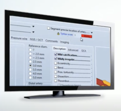
Contributing to this standardization of reporting between cardiologists, and to easing the reporting time burden, is something ScImage calls “evidence-based reporting.”
In the dark era (and in many labs to the present day) cardiologists would make subjective interpretations of the size of the cardiac chamber, the function of the ventricles and the severity of valvular disease. This created huge inconsistencies in interpretation.
In my lab we emphasize precise measurement of these crucial cardiac parameters using the sonographer-generated measurements, and a range that we specify for each parameter using American Society of Echocardiography (ASE) guidelines. The report is automatically populated with the corresponding appropriate statement. For example, the echo I was reviewing had a left atrial volume index of 35 (just over the upper limits of normal). “Mild left atrial enlargement” was already present in my report, along with the correct description of left ventricle (LV) and right ventricle (RV) size and function as well as right atrium (RA) size.
Thus, with a few clicks, I had completed the report between bites of the truffle chicken chanterelle flatbread (beurre fondue, grilled chicken, chanterelle mushrooms, parmesan, truffle oil) the Eternal Fiancée had just ordered.
As we were settling the bill, I received a page — I was being consulted on a 92-year-old man (Mr. Card Infarct) in the ER with chest pain and elevated troponins. In the dark era, the only information I would have available to me would be what the nurse or ER doctor on the phone told me. If they were ill-informed or English-challenged, a trip to the hospital likely would be necessary.
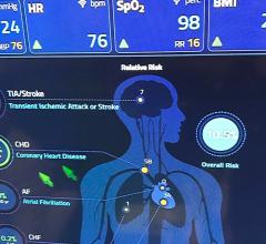
In 2019, fortunately, while sitting at the Oceano bar, I can review my partner’s office notes on Mr. Infarct and review everything that has been done on him in our hospital’s system, including a coronary artery bypass graft (CABG) in 2015, a stent in 2017, and an implantable cardioverter defibrillator (ICD) in 2017. Since our cardiology PACS has all modalities viewable in one list from one login, it is a very simple matter for me to review Mr. Infarct’s last catheterization, his last echo and compare his prior ECGs to the current one. Within five minutes I pretty much am armed with everything I need to know about Mr. Infarct in order to best handle his case. When I call the ER, I can easily process the information the ER doctor gives me about his presenting signs and symptoms and develop a plan.
Life for this on-call cardiologist is good. It’s more normalized here in the digitally enlightened era. I’m no longer shackled to the hospital. And I can enjoy excellent appetizers in a convivial environment without (excess) guilt or anxiety.
Transoceanically Yours — Anthony Pearson, M.D.
PICOM365 by ScImage integrates all cardiac imaging disciplines’ data — Echo, Cath, Vascular, Nuclear Medicine, Holter, Stress and ECG studies — in one enterprise PACS solution accessible from anywhere, any time. Learn more at www.scimage.com.


 November 06, 2025