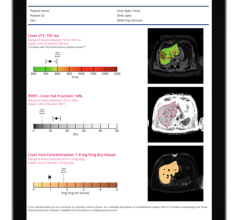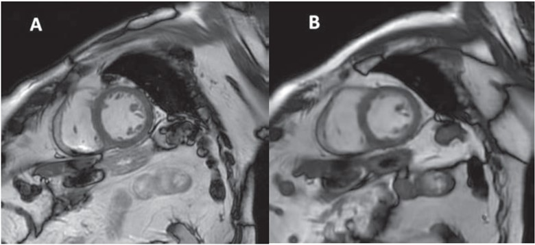
85-year-old patient with ischemic heart disease, who underwent cardiac MRI at 3 T.
A. Short-axis image from standard cine balanced SSFP breath-hold sequence.
B. Short-axis image from free-breathing accelerated cine sequence with deep-learning reconstruction. Images show normal appearance of left ventricular myocardium.
February 12, 2024 — According to the American Journal of Roentgenology (AJR), free-breathing cine-deep learning (DL) may yield reliable left ventricular ejection fraction (LVEF) measurements in patients with ischemic heart disease (IHD) unable to repeatedly breath-hold.
“Free-breathing cine-DL sequence, in comparison with standard breath-hold cine sequence, showed very small bias for LVEF measurements and better subjective quality,” wrote David Monteuuis, MD, from the department of radiology at Amiens University Hospital in France. “Cine-DL yielded greater left ventricular volumes.”
David et al.’s AJR accepted manuscript included patients undergoing 1.5-T or 3-T cardiac MRI for evaluation of IHD (March 15–June 21, 2023). Examinations included an investigational free-breathing cine short-axis sequence with DL reconstruction (cine-DL). In blinded fashion, two radiologists (R1, R2) independently assessed LVEF, left ventricular end-diastolic volume (LVEDV), left ventricular end-systolic volume (LVESV), and subjective image quality, for cine-DL sequence and standard breath-hold balanced SSFP sequences; R1 assessed artifacts.
Ultimately, in patients with IHD, the free-breathing cine sequence with DL reconstruction showed very low bias for LVEFmeasurements (R1: 0.4%; R2: 0.7%) and better subjective image quality (R1: 2.3±0.5 vs 1.9±0.8; R2: 2.2±0.4 vs 1.9±0.7) in shorter acquisition times (0.6±0.1 vs 2.4±0.6 min).
“This free-breathing cine-DL sequence could be particularly useful in the evaluation of patients with dyspnea who are unable to perform repeated breath-holds,” the AJR authors added.
For more information: www.arrs.or

 May 14, 2026