September 1, 2010 - Two devices in one is one of the mega trends in imaging diagnostics. The spectrum ranges from a combination of positron emission tomography (PET) with magnetic resonance imaging (MRI) or computer tomography (CT) to a combination of ultrasound with endoscopy. At MEDICA 2010, World Forum for Medicine - International Trade Fair and Congress, to be held from Nov. 17–20, 2010 in Düsseldorf, Germany, medical technology companies and congress speakers will focus on innovative hybrid procedures such as endosonography and its application in the daily clinical routine. Use of these so-called hybrid procedures is increasing, primarily on cancer patients.
The idea is obvious: Combine two diagnostic procedures in a single examination in order to obtain the information from two procedures, not just from one imaging method, more easily and with less stress for the patient. This is made possible by these hybrid procedures, in which two different imaging methods are combined in a single comprehensive technical system.
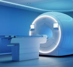
One example of a special hybrid system is the whole-body PET / MRT scanner, equipped with the latest Philips technology, which was installed at the University Hospital of Geneva this year. However, it will still be used primarily for research purposes. This system combines two procedures which were not combinable up to now, namely magnetic resonance tomography (MRT) and positron emission tomography (PET). The combination of MRT and PET allows the spatial structures and the metabolism activity of the organs to be displayed in a single image. This is of particular interest in cancer treatment, because tumor cells often require more energy than the healthy cells in adjacent tissue. This is important for initial diagnosis, surgery planning and verifying how successfully the tumor was treated.
“The combination of anatomical imaging by MRT and the metabolism through the applied, radioactively marked tracer for PET will assist in monitoring and prediction as well as supervising the treatment of cancer patients and will provide more precise information on how the patients respond to treatments. We also believe that hybrid imaging will also have more potential in other areas, particularly in cardiovascular imaging as well as neurology,” explains professor Osman Ratib, chief physician at the Clinic of Radiology and Nuclear Medicine of the University of Geneva.
Currently, both MRT and PET examinations are performed separately, often over several days. Then the images are superimposed on the processing console. Consequently it is very difficult to match the images precisely, as the patient is never in exactly the same position during the examinations and the alignment of the respective scanners does not always correspond precisely. Large diagnostic devices that combine PET and computer tomography have been in use for years as PET/CT devices. In contrast, the combination MRT and PET was thought to be virtually unfeasible. “Up to now, the magnetic field of the MRT prevented the proper functioning of the PET scanner and created artefacts,” says Professor Ratib. These problems have been solved in the new whole-body PET / MRT scanner. With the hybrid device, the two scanners are located in a room opposite to each other. In between is a rotating table, so that PET and MRT can take place one after the other without the patient changing position. “We chose three particular areas in which we expect a significant improvement,” states Professor Ratib. These are, firstly, patients with head/neck tumors where the evaluation of tumor recidivation with conventional imaging is often very difficult due to the frequently very radical surgery. The PET / MRT will also be used for prostate cancer, where the primary aim is the early recognition of recidivation, and for breast cancer, where the procedure will help to improve the differential diagnostics.
Significantly less examination stress
Radiologists at the University Hospital of Tübingen have had a combined PET MRT system for a year already. Professor Claus D. Claussen, Medical Director at the Department of Radiology, remarks: “For our patients, this means that in future there will be significantly less examination stress because the examination time can be considerably reduced and the exposure to radiation will be eliminated. This technology allows earlier recognition of tumors and metastases, they can be characterized more clearly, and their location relative to the organs more definitely established. The expected result will be earlier and more targeted therapy.” The University Hospital has already completed important steps in the development of PET-MRT for the head. Now the challenge for scientists is to adapt the imaging technology for the head, which is relatively small, to the whole body. The PET-MR for brain imaging was developed in cooperation with Siemens.
Researchers in Jülich expect to recognize brain diseases such as Alzheimer’s in the very early stages with a PET plus a strong MRT of 9.4 tesla (that is almost 200,000 times stronger than the earth’s magnetic field) – a technology also developed by Siemens. The device simultaneously records the tissue structure and biochemical processes in the brain. The director of the Institute for Neurosciences and Medicine, Professor Jon Shah, expects a resolution of under 0.1 millimetres. Researchers also expect advances in basic research, for instance with respect to addiction and headache. However, improved diagnosis does not mean immediate improved therapy. It may take years until new therapeutic agents developed on the basis of this research can be tested.
Combination of MR and ultrasound
With MRgFUS and MR-Touch, the US company GE Healthcare has two hybrid procedures in its range of products. MRgFUS is a combination of MRT and highly focused ultrasound for myoma therapy. MR-Touch is a special elastography procedure that allows visual scanning of organs. For centuries, physicians have relied on their sense of touch when examining their patients, for instance when feeling for lumps in the breast or palpating the liver in order to recognize liver fibrosis. However, not all the organs can be reached like that. MR-Touch provides the solution. The process utilizes a combination of low-frequency sound waves and MR technology to measure the elasticity of the tissue. This produces an elastogram, a color-coded anatomical image of the firmness of the liver tissue. The process comprises three steps. First, sound waves (between 40 and 200 Hz) are generated in the body by means of an MRT-compatible generator. In the second step, an image of these sound waves is created with a special MR imaging sequence. In the third and last step, these data are processed and an elastogram is created which shows the relative firmness of the tissue in the area being examined.
The advantage of endosonography (EUS, endoscopic ultrasound) is also the combination of two imaging procedures, in which the organs are not examined from the outside through the skin, but from the inside. The ultrasound head directly contacts interior surfaces such as the mucosa of the oesophagus by means of an endoscope. In contrast to conventional ultrasound examinations through the skin, the advantage of this procedure is that the target organ is closer to the ultrasound head, therefore allowing it to be displayed more precisely or actually displayed at all. A balloon filled with water is often placed on the tip of the endoscope in order to enhance the display. This improves conduction of the ultrasound waves to the tissue as well as improved reflection. Different devices are used, for instance those made by Olympus, depending of the area of application. Miniprobe systems are related to endosonography. These are instruments that can be pushed through a biopsy channel, for instance. However, the radius of the penetration depth of the ultrasound is smaller. Miniprobe endosonography is particularly suitable for such procedures as the targeted examination of a polyp or to estimate the depth of a malignant tumor.
Endosonography gains in significance
Endosonography “allows the observation of almost microscopically precise sectional images of the intestine wall,” explains Dr. Elke Burmester of Sana Clinics in Lübeck. The examination itself is relatively simple and not very stressful for patients. The significance of endosonography in imaging diagnostics, above all for colorectal diseases, has increased in recent years and it is now considered an important diagnostic building block. On the one hand this applies for assessment of the continence organ, anorectal abscesses and fistulas. On the other hand, with respect to cases of rectal carcinoma endosonography is now an obligatory component of the current S3-guidelines (S3 = highest guideline classification issued by Germany’s Association of Scientific Medical Societies AWMF). Determination of the tumor infiltration depth and evidence of lymph node metastases is very precise. The hybrid procedure allows exact therapy control with tumor follow-up, through which it is possible to recognize and precisely puncture suspicious findings.
This procedure is also an option in the diagnosis of tumors in the area of the pancreas and in cases of unclear cholistasis. According to Burmester, the results of EUS in pancreatic procedures are rather unsatisfactory. In addition, endosonography is particularly sensitive in the diagnosis of neuroendocrine tumors of the pancreas. It is even indispensable for the diagnosis of insulinoma for pre-operative planning.
At MEDICA 2010, the state of modern tumor therapy is the topic of several congress events held at the Congress Center South.
For more information: www.medica-tradefair.com

 May 14, 2026