News | Cardiac Imaging | May 12, 2021

Using Contrast MRI After a Heart Attack Could Increase Survival

According to the British Heart Foundation, heart and circulatory diseases cause more than a quarter (27 percent) of all deaths in the UK, which equates to more than 160,000 deaths each year — or one death every three minutes.

May 12, 2021 — According to the British Heart Foundation, heart and circulatory diseases cause more than a quarter (27 percent) of all deaths in the UK, which equates to more than 160,000 deaths each year — or one death every three minutes.

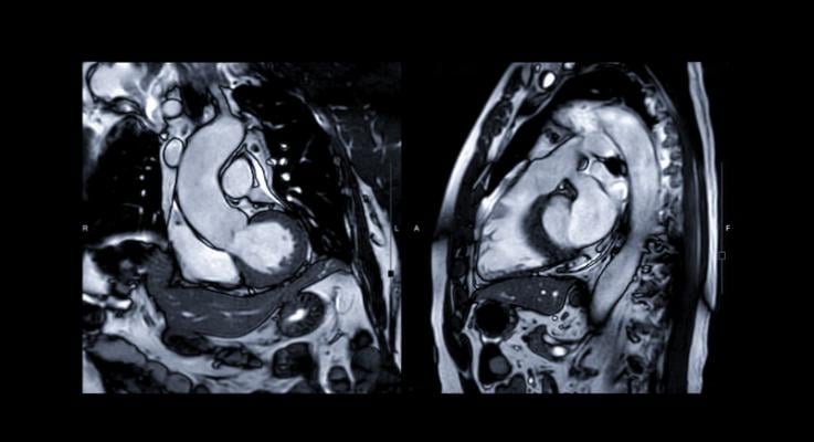
The research, published in the top science journal Advanced Science, found that injection of the trace mineral manganese could enhance MRI scans so that they provided more accurate details of heart function than traditional MRI methods.

These findings, if confirmed in human subjects, could have major implications for the treatment of heart attack patients. The findings could also be of great use in the preclinical evaluation of treatments for patients who suffer from cardiac ischemia — a reduction in blood supply to the heart muscle that could lead to cardiac arrest.

The study also suggests that if manganese-enhanced MRI is performed within the first few hours of a heart attack it could be used to determine the optimal treatment regime for individual patients — helping to regulate changes in the cardiac muscle and thereby further improving survival chances. Findings were evaluated by examining the infarct size and blood supply at three key intervals: one hour, one day and 14 days after a myocardial infarction was induced.

Patrizia Camelliti, M.D., Principal Investigator and Senior Lecturer in Cardiovascular Science at the University of Surrey, said: "Magnetic resonance imaging (MRI) is increasingly used to diagnose and give information on heart conditions. This research using mice allows us to measure the health status of the heart muscle rapidly after a heart attack and could provide important information for optimising treatments in patients."

For more information: http://www.surrey.ac.uk


Related Content

News | Cardiac Imaging

April 23, 2024 — CDL Nuclear Technologies, a pioneer in advanced diagnostic solutions, is proud to announce the launch ...

Home April 23, 2024
Home
Feature | Cardiac Imaging

Cardiac positron emission tomography (PET) is growing in popularity among cardiologists because it provides the ability ...

Home March 05, 2024
Home
News | Cardiac Imaging

PLEASE NOTE: This webinar has been postponed to a later date. A new date will be posted in the coming days. On March 25 ...

Home February 29, 2024
Home
News | Cardiac Imaging

February 28, 2024 — Royal Philips, a global leader in health technology, announced major enhancements to its Image ...

Home February 28, 2024
Home
News | Cardiac Imaging

February 12, 2024 — According to the American Journal of Roentgenology (AJR), free-breathing cine-deep learning (DL) may ...

Home February 12, 2024
Home
News | Cardiac Imaging

November 16, 2023 — Cardiovascular diseases rank among the top causes of death across the world, and cardiac ...

Home November 16, 2023
Home
News | Cardiac Imaging

September 21, 2023 — Declines in cardiovascular procedure volumes observed early in the COVID-19 pandemic greatly ...

Home September 21, 2023
Home
News | Cardiac Imaging

July 11, 2023 — Hyperfine, Inc., the groundbreaking medical device company that created the Swoop system, the world’s ...

Home July 11, 2023
Home
Feature | Cardiac Imaging | By Matthew Jay Budoff, MD, FACC, FAHA

Coronary computed tomography angiography (CCTA) is cheaper, easier to use and its results are easier to understand than ...

Home July 10, 2023
Home
News | Cardiac Imaging

June 29, 2023 — According to an accepted manuscript published in ARRS’ own American Journal of Roentgenology (AJR) ...

Home June 29, 2023
Home
Subscribe Now