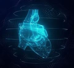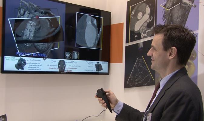
Technology developed by Teistler Image allows one-hand operation of a game controller to intuitively slice through any axis on a CT scan of a heart
November 20, 2013 – Video game technology is being applied to imaging by German researchers, who will be presenting their findings at RSNA 2013. Xbox in the operating room and the use of video game controllers in visualizing organs and tumors, an ultra-sensitive MRI technology for early detection of cancer and a one-stop stroke management technology are among advances coming from scientists affiliated with the German Federal Ministry for Education and Research (BMBF):
- Xbox has a place in the operating room: Technology built on the Xbox Kinect lets surgeons pull up medical images, switch to a different image or zoom in on an image, all without touching a thing, making it useful even if they are scrubbed for surgery. New time-of-flight 3-D camera gesture navigation gives surgeons control without physical contact with the technology.
- Video game controller enables remote control exploration of organs, tumors: Using an off-the-shelf game controller, radiologists can easily and quickly navigate through 3-D images of a tumor or beating heart from any angle or any direction. The software generates special 3-D or 4-D versions of computed tomography (CT) or magnetic resonance (MR) images, which the radiologist can manipulate and explore by twisting and turning the game controller – instead of a mouse, touchpad or keyboard — providing more flexibility and enabling more viewing option. The technology developed by Teistler Image allows one-hand operation of a game controller to intuitively slice through any axis on a CT scan of a heart or other organ.
- Highly sensitive + specific molecular imaging for early disease detection: A new technique called hyper-CEST for MRI can capture highly specific images in less than two minutes and detect cellular changes in human tissue far too small to be visible using standard MRI. This advance holds promise for earlier diagnosis of diseases such as pancreatic cancer, which typically are not detected until they are late stage. The technology uses harmless xenon gas and produces images that would require 1,100 years to make using traditional methods.
- Faster stroke diagnosis; better treatment: Doctors will be able to quickly and precisely image the entire brain and its blood flow to determine whether a stroke is being caused by an aneurysm or a blockage – and potentially treat the patient in the same room — using a new multimodal functional imaging system that enables “one-stop stroke management.” Being able to diagnose and treat stroke more quickly would help reduce damage and save lives.
- Visualized tumor tracking over time enables easier assessment; quicker action: New software can analyze all of the many images taken over time of a patient’s tumor and quickly show how it is responding to treatment. By tracking the tumor and speeding up analysis, doctors can assess response to treatment and change therapy if needed. The first and only software of its kind, mint Lesion analyzes all MR, CT and other imaging and test data, reporting findings in a single record. The program can be used to track groups of patients as well as individuals, making it useful for clinical trials, and providing more objective data.
Learn more by visiting the BMBF booth 2965 at RSNA 2013.
For more information: http://www.bmbf.de/en/

 May 14, 2026