June 9, 2014 — At the 2014 Annual Meeting of the Society of Nuclear Medicine and Molecular Imaging (SNMMI), June 7-11, at the St. Louis Convention Center, Siemens Healthcare will debut syngo.via for molecular imaging — a reading solution that transforms large amounts of data into comprehensive results, allowing for organ-based reading, reference-based quantification and evidence-based reporting of disease. With syngo.via for molecular imaging, physicians are better able to focus on reading and can more easily produce high-quality diagnostic reports. Also at this year’s SNMMI, Siemens will move from innovation to results, demonstrating the clinical adoption and efficacy of the two groundbreaking molecular imaging systems it introduced at last year’s SNMMI annual meeting — the Biograph mCT Flow positron emission tomography/computed tomography (PET/CT) system with continuous FlowMotion technology; and Symbia Intevo4, the first system to completely integrate combine datasets from single-photon emission computed tomography (SPECT) and CT.
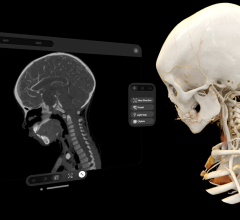
syngo.via for Molecular Imaging
Physicians expect clinical applications software to help better understand complex diseases at a functional level. The new syngo.via for molecular imaging provides unique and automated tools to visualize, measure, and report disease.
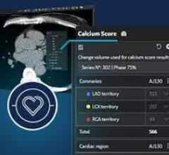
Now, physicians can immediately review all available information and perform organ-specific readings. For multiple time-point cases, prior studies are all registered with each other, even when acquired on multiple sources. The new Siemens-exclusive EQ•PET algorithm harmonizes quantification of PET data based on National Electrical Manufacturers Association (NEMA) references. It allows standard uptake value (SUV) comparison in longitudinal patient exams for determination of cancer manifestation and change, independent of scanner make, model,or reconstruction algorithms. Neurology quantitative tools provide comparable results, which could potentially improve reader agreement for Alzheimer’s disease evaluation. Cardiology quantitative tools improve therapy risk management of coronary artery disease with diagnostic blood flow quantification. Finally, with syngo.via structured reports, molecular imaging physicians will be key contributors to confident therapy decisions of the multi-disciplinary patient management team.
Biograph mCT Flow: One Year Later
At SNMMI 2013, Siemens first detailed how its Biograph mCT Flow overcomes the limitations of conventional bed-based PET/CT with FlowMotion, a new technology that moves the patient smoothly through the system’s gantry while continuously acquiring PET data. Biograph mCT Flow with FlowMotion advances routine image quality to a new level by enabling imaging protocols based on the organ’s need. FlowMotion expands accurate and reproducible quantification in all dimensions for precise disease characterization in therapy monitoring, while enabling physicians to offer as low as reasonably achievable (ALARA) dose to every patient.
A year after the introduction of FlowMotion, the PET Center at the University of Michigan Hospitals in Ann Arbor is routinely demonstrating how high-resolution FlowMotion studies can help physicians discover tiny lesions in the head and neck that might otherwise elude detection. “We [scan] our head and neck cancer patients with FlowMotion because it permits us to very efficiently acquire better images,” says Kirk Frey, M.D., director of the PET Center. “We believe this gives us more accurate nodal staging. Our sense is that FlowMotion provides an accurate and sensitive acquisition.”
Pioneer users of FlowMotion technology are also using it to perform whole-body imaging — either routinely for oncologic applications or as part of research projects. Keio University School of Medicine in Tokyo began using FlowMotion routinely in February, utilizing it primarily in oncology. “FlowMotion allows us to visualize the tiniest lesions in head and neck cancer patients,” says Koji Murakami, M.D., Ph.D., head of the division of nuclear medicine, department of radiology. “For these patients, we perform precise imaging using slow table speeds, followed by a faster table speed for the pelvis and extremities, which have a lower probability of metastases.”
Respiratory gating is an important part of scans involving suspected thoracic and abdominal metastasis, yet until recently, Murakami and his colleagues had no choice but to conduct a separate respiratory gated scan for these patients in addition to a whole-body, stop-and-go scan. Biograph mCT Flow with FlowMotion resolves this problem. “The merit of FlowMotion,” he says, “is that we can vary the acquisition time and integrate respiratory gating into a single exam.”
Symbia Intevo: One Year Later
At SNMMI 2013, Siemens debuted Symbia Intevo, the world’s first xSPECT system, which combines the high sensitivity of SPECT with the high specificity of CT. Completely integrating data from both modalities, Symbia Intevo generates high resolution and, for the first time ever, quantitative images for SPECT. xSPECT reconstructs both the SPECT and CT portions of the image using the high-resolution CT frame of reference for precise, accurate alignment that facilitates the extraction and deep integration of medically irrelevant information.
At the German Federal Armed Forces Hospital in Ulm, Symbia Intevo with xSPECT made an immediate impact upon its arrival in March. The system delivers xSPECT images that localize metabolic hot spots so lesions on the bone can be differentiated from those in the surrounding soft tissue. “They look like CT images — the outlying border is extraordinary; the brilliance is very good,” says Burkhard Klemenz, head of the hospital’s nuclear medicine department. “Now, for the first time, I look at the xSPECT Bone [4] images, and I talk to my colleagues only about those images; it’s because the images are so convincing. We can recognize disease localization now with xSPECT Bone.”
For more information: www.siemens.com/healthcare

 December 23, 2025