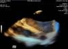
Siemens' AcuNav V 3-D intracardiac echo (ICE) catheter offers detailed, live 3-D images of the interior of the heart. This video shows an example of the catheter imaging the pulmonary vein. The technology may play a role in better guiding transcatheter electrophysiology (EP) ablation procedures. The technology was shown as a work-in-progress during ACC 2012.
Videos
VIDEO: Emerging Trends in Cardiovascular Imaging
Cardiac Imaging | January 23, 2023
Artificial intelligence and general consolidation were two top cardiology trends at RSNA22. ITN/DAIC spoke with Val Kapitula, partner, Paragon Consulting Partners LLC, about some of the trends he is seeing emerge in this field.
Related content:
RSNA 2022 Theme in Action: Empowering Patients and Partners in Care
Photo Gallery of Radiology Technology, Highlights from RSNA 2022
RSNA 2022 Day 4: Back to Basics on AI, ABUS Training, and ML in RO
RSNA 2022 Plenary Speaker Omary Urges Radiologists to Support Patients, Communities and the Planet
RSNA 2022: Innovative Solutions on Display, Leadership Addresses Value of Imaging
RSNA 2022 Day 2: Focus on Breast Screening & AI, Future of Patient Care
Cardiac Imaging
Ultrasound Intra-cardiac Echo (ICE) | April 06, 2012
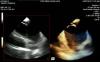
Siemens' AcuNav V 3-D intracardiac echo (ICE) catheter offers detailed, live 3-D images of the interior of the heart. This video shows an example of the catheter imaging a transseptal puncture. This new ICE technology may help better guide these punctures, which are routinely used in catheter ablations and transcatheter left atrial appendage (LAA) occluder delivery. The technology was shown as a work-in-progress during ACC 2012.
ACC | March 30, 2012

Diagnostic and Interventional Cardiology Editor Dave Fornell discusses trends and shares his choices of the most innovative technologies shown on the floor of the American College of Cardiology (ACC) 2012 Scientific Session, held March 24-27 in Chicago.
A couple of key trends were evident on the show floor included:
• New technology to support trans-aortic valve replacement (TAVR);
• Launch of new cardiovascular image and information systems (CVIS) to support healthcare's adoption of proposed Stage 2 meaningful use (MU) requirements;
• Balloon-inflatable TAVR/EVAR introducer sheath;
• 3-D intra-cardiac echo;
• Mobile angiography system for hybrid ORs;
• Chocolate for heart health.
For more information: www.DIcardiology.com
Cardiac Imaging | December 30, 2011

DAIC editor Dave Fornell explains some of the most innovative cardiovascular imaging technologies showcased by vendors at the Radiological Society of North America (RSNA) meeting in December 2011.
Ultrasound Imaging | December 16, 2011
Toshiba (Canon) unveiled its Aplio 500 ultrasound system at RSNA 2011, , which offers a unique 3-D fly-through imaging capability. The system takes the image dataset and processes it to create a cine loop fly-through of any hollow, fluid-filled blood vessel, duct or organ. The example in this video is of a blood vessel in the liver. The capability and image quality is similar to what is seen in a virtual colonoscopy created from CT datasets. The technology was highlighted in our editor's choice for most innovative new technologies at RSNA 2011. The future applications of this technology may include 3-D ultrasound navigation aids for vessels in the cath lab.
PET-MRI | June 27, 2011

Three companies showed different versions of a combined positron emission tomography (PET)-magnetic resonance (MR) (PET-MRI) system during the Society of Nuclear Medicine (SNM) 2011 annual meeting. Representatives from Siemens, Philips and GE Healthcare explain how their systems work and how PET-MR may be used as a new modality to show both physiologic and anatomical information.
Each company took a different approach to how they create PET-MRI images. Siemens integrated both modalities into one gantry. Philips uses two gantries with a table that moves between the two that maintains patient alignment for fusion imaging. GE Healthcare uses a cot that can move between the MR and PET rooms and fits both systems to maintain alignment and does not require buying a new decicated scanner.
Nuclear Imaging | June 27, 2011

Society of Nuclear Medicine (SNM) President George Segall, M.D., chief of the nuclear medicine service at the VA Palo Alto Health Care System, and is a professor of radiology and professor of cardiology (by courtesy) at Stanford University School of Medicine, offers insights into the trends he saw at the society's 2011 annual meeting.
Trends in nuclear imaging include the creation of PET/MRI systems, use of time of flight (TOF) imaging, new technqiues to image amyloid plaque in Alzheimer's Disease, and the movement toward multimodlaity imaging rather than radiologists specializing in justy one modality.