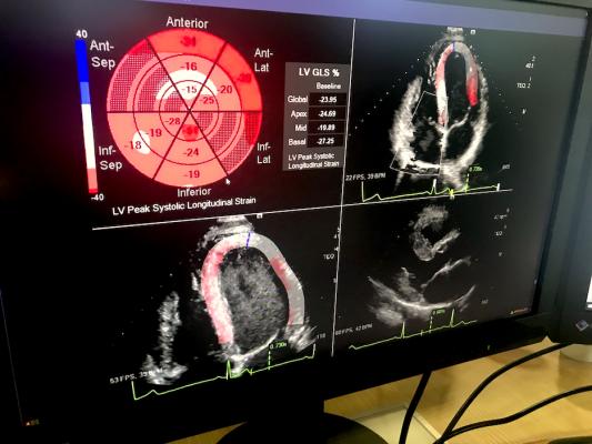
An example at HIMSS of deeper third-party software integration in enterprise imaging platforms was Siemens Healthcare showing a new, deep integration with echocardiography strain imaging analysis provider Epsilon Imaging. The integration eliminates the need for a separate login and the Epsilon software is now just a tool option available in the syngo system and it carries over data and images directly into the echo report.
Taking advantage of new technology advances, several radiology PACS, enterprise imaging and cardiovascular information system (CVIS) vendors are releasing new, completely revamped platforms. This was a trend across vendors we visited at the the 2021 Healthcare Information Management Systems Society (HIMSS) in-person meeting in August.
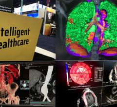
The newer platforms are increasing speeds to access images and report, better flow and integration with vendor neutral archives, use of zero-footprint image viewers for both radiologists and referring physicians, and deeper integrations of artificial intelligence (AI) and third-party applications so they are seamless in the workflow.
"About 90% of new system installations are going to be cloud-based solutions," said Brad Levin, Visage Imaging general manager, North America and global head of marketing. He said the trend for enterprise imaging systems and PACS is clearly going moving to cloud based solutions because it solves several issues for hospitals.
Use of cloud-based systems also help with easier integration with large hospital electronic medical record (EMR) systems. These vendors explained they can now easily integrate with most of the larger EMR vendors, most notably Epic, Cerner and Allscripts.
Some vendors are also moving to larger cloud data storage platforms, such as Google Cloud and Amazon Web Services (AWS). The value in the larger platforms is that they can scale immediately for any since healthcare enterprise and the hospitals no longer need to maintain and upgrade servers. But the biggest asset is said to be the higher level of security with these mega cloud server companies. They have large, dedicated IT teams that monitor data security 24/7.
"The cloud is bringing a lot of scalability and physicians can focus on patients rather than on IT concerns," explained Tracy Byers, Change Healthcare general manager for enterprise imaging.
An example of one of the new systems introduced at HIMSS is GE Healthcare's new Edison Cloud PACS. It leverages AI and fully integrates it into the radiology workflow. It also has an updated structure to allow better operation in the cloud. It is replacing GE's older radiology IT systems.
Edison allows users to easily customize their own work lists. The work lists show if anyone is currently in a study, who they are and if they are dictating. Users can also go through the list and reserve the studies that pertain to their area of expertise. A messaging system allows radiologists to send text notes to others to see if they can go into a study or other communications. Users also can create and pin groups for specific subspecialty reads.
The new PACS cloud is hosted by AWS. This large cloud provider has a dedicated IT security staff that works 24/7 to maintain data security, which offers more resources than most hospital IT teams.
Siemens introduced its new syngo Carbon enterprise IT platform that will serve as an umbrella to allow a single access view for all patient data across an enterprise. The cloud-based platform includes image and data management, an image viewer, and a storage container to feed in enterprise data from all departments. Similar to GE's Edison system, Siemens said Carbon also is optimized so AI functions can be added to enable for predictive workflow and an array of new technology applications.
Users will be able to call up syngo tool sets such as advanced visualization tools from anywhere in the enterprise. Auto quantification functions will be incorporated into reports. AI apps for various functions also will be fully integrated now, rather than sitting on top of the PACS or CVIS platforms as third-party interfaces. The system offers a zero-footprint viewer.
Visage Imaging introduced new cloud PACS solution, consolidating reporting, a multi-modality viewer, storage, and advanced visualization tools into one location and accessible from anywhere with an internet connection. The system is cloud native, so it is optimized for cloud operation, rather than an older system retrofitted to save and retrieve data from the cloud.
Change Healthcare introduced Stratus Imaging PACS at HIMSS, its first cloud-native offering. It is a comprehensive cloud software as a service (SaaS) solution for radiology practices. The platform will replace older generation systems offers by Change and its acquired former McKesson products.
Change said cybersecurity is a major concerns by hospitals and radiology practices and moving to the cloud offers a mach more secure platform because the cloud providers have large teams monitoring and patching security issues.
On the cardiology side, Change is working on a completely new hemodynamic system, which will replace its older version that is missing parameters needed for new structural heart and EP procedures, and it was found to not be scalable between ambulatory surgical centers and large IDNs. The new demo system will be introduced in 2022.
A cardiology trend noted by Change Healthcare is the need to integrate easily with Epic and other larger EHR systems. Often hospitals use Epic Cupid for basic cardiology information, but they want the much more detailed CVIS reporting workflow offered by third-party vendors like Change.
Fujifilm recently released its new Synapse 7X enterprise imaging platform. The vendors acquired Hitachi's health IT segment and integrated its Vidastar cardiology reporting and image viewer and the Hitachi analytics models into Synapse. The cardiac segment is also tailored for specific pediatric cardiology reporting.
Advanced visualization vendor Vital Images (now Canon) has greatly evolved over the past few years by leveraging the ability of its Vitrea software to interoperate with various vendors. Canon has turned this technology into a full service enterprise imaging platform. The platform is now used by Ochsner Health in New Orleans, integrating cardiology, radiology, and endoscopy. It allows customization of hanging protocols, such as adding automated MPRs rather than setting this on the scanner or creating them in a 3D lab. It also offers a simplified web URL based viewer for referring physicians to access the images and reports.
Vitrea Connect is the system’s data orchestration layer. Rather than converting data formats into something easy for the system to ingest and store, such as a proprietary format, it stores everything in its native format. When images are opened, they are opened in the original format. This allows disparate data to be combined into the system’s VNA and data is pulled from one source. This can also help normalize data to make later transfer into another archive possible. It also fully integrates AI apps, rather than adding them as a separate layer or module.
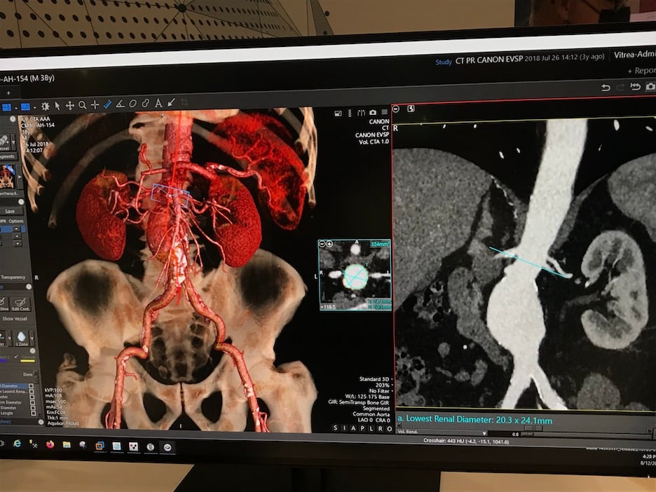
Canon's Vitrea PACS data orchestration layer uses the original data formats for images. When images are opened, they are opened in the original format. The platform allows multimodality images and advanced visualization reformatted images to be displayed together in the same viewer.
Speed Has Become Key for PACS and CVIS Platforms
Another benefit of the newer generation web-based, cloud platforms is they offer much faster connection speeds. Large imaging studies can now be opened in less than a second or two, rather than waiting for long before the images appear and can be read.
Change Healthcare and Visage Imaging both used examples in their booth demos of 3D mammography tomosynthesis studies. In both cases, the vendors demonstrated they could open all the images in the study, from the cloud in less than 3 seconds. In the case of Visage, the exam was opened in Las Vegas from a remote server located in Iowa. This also included linked prior exams for the same patient.
Adapting Workflow to Better Fit Needs of Physicians
Adapting workflow to better fit radiologists was a theme explained by several vendors at HIMSS. Vendors displayed various workflow improvements on their systems, including automated hanging protocols, auto fetching and display of prior or related images of the same anatomy, and reducing clicks for various operations.
Change Healthcare demonstrated an example of this with a feature called 3-D Rotate that radiologists can click on and add a center line on any axis in the anatomy of a CT scan. They can then scroll through the dataset along that line rather than the usual fixed planes on standard views. It does not require the radiologist to use a different viewer or separate program, allowing them to continue working in one viewer to save time.
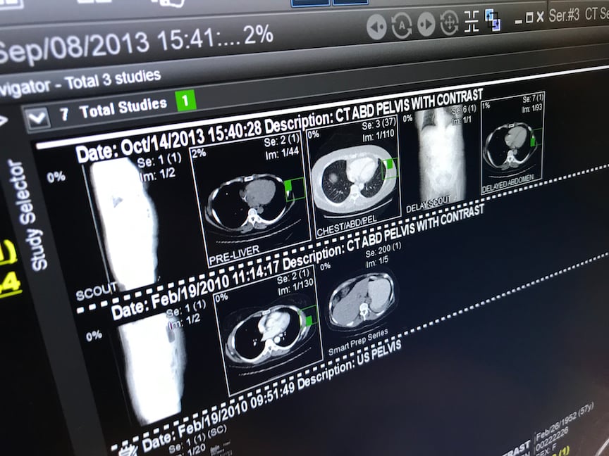
An example of an automated PACS hanging protocol that brings in relevant prior images displayed by Infinitt. The system pulls in comparable prior exams of the anatomy in the current exam. It shows a prior exam timeline at the top of the page and allows quick a quick review. The user can click on the preview images to take a peek so see if it is relevant before opening the entire exam. The images also sync to show the same slices of anatomy. As the radiologist scrolls through the current exam, the system will also roll through the prior exam at the same time. This offers a sizable time savings in reading the exam and prior. The hanging protocols also can be set to call up either MIP or MPR images automatically, based on the radiologists preferences.
To speed mammography reads on the GE Edison platform, as a patient's exam is opened, the system automatically fetches all prior mammograms. These are presented with the most recent prior side by side with the current exam. The radiologist can just click use one click at a time to flip through the other prior exams, which are numbered so they can access more information from if needed.
GE also showed a small tweak to improve flow of images by creating a small icon on the various exams and priors called up. A small green box on the thumbnails of the exams show where theses are located in the multiple screens the radiologist currently has open. They can click on that exam they have interest in and it auto populates they main screen for closer review.
While having a large number of tools for image analysis and reporting may seem like a good things, Agfa said some hospitals, especially smaller center, are asking for easier customization to turn off many of the advanced features so they just have the common tools they need appearing in the main menus. Agfa altered its enterprise imaging system to allow easier customization, including a variety of choices with image hanging protocols.
GE also showed a small tweak to improve flow of images by creating a small icon on the various exams and priors called up. A small green box on the thumbnails of the exams show where theses are located in the multiple screens the radiologist currently has open. They can click on that exam they have interest in and it auto populates they main screen for closer review.
Seamless Integration of Artificial Intelligence Into PACS and CVIS is Now a Trend
About three or four years ago, most health IT vendors realized they did not have the resources to develop in-house AI applications for every conceivable use. As a result, many began to partner with AI start-ups to offer their AI technology after they gained regulatory approval. The primary model developed was an App Store-like environment, where hospitals could pick and choose which AI apps they wanted and there was some level of integration guaranteed with the App Store vendor's primary IT systems.
However, vendors are now realizing that radiologists and cardiologists will not use AI apps if they are cumbersome, requiring extra logins, extra steps to access the AI, or the AI data being saved someplace separate from the rest of the past data. For this reason, many PACS and CVIC vendors are now looking beyond an App Store approach to full, seamless integration with the AI as if the app was developed as part of the system. This eliminated the need for interfaces, separate documentation, separate images, separate logins or opening software that sits as a separate layer on the main reporting or image viewing applications.
Companies that are now taking this approach include Agfa, Vital, GE Healthcare and Fujifilm.
"It is too hard to use stand-alone apps. You need to have a solid integration into the workflow," explained Bob Craske, Agfa's director of enterprise imaging, radiology and clinical application.
Radiologists have become very accepting of AI in the past couple years as these algorithms gain clearance from the FDA. Craske said the discussion has moved from how the AI works to just having conversation on how this will help them in improve workflow. The areas where the vendor has seen most interest has been AI for chest CT lesions, chest X-ray bone removal, and indices of areas of concern identified by the AI to provide a backup review for the radiologists.
Another trend Craske has seen with AI is academic centers asking for a way to switch off the AI for some users, such as newer residents, so they do not become reliant on the technology.
Siemens is creating a series of AI care pathways on its enterprise imaging platform, where the AI pulls in all the relevant data and prior exams for a patient just for a specific condition. This includes a dash board view of a cancer patient's data relevant just to their cancer. It also can be shown as a timeline flow chart, where the AI will suggest next steps for diagnosis or treatment based on the latest guidelines. The system also uses predictive analytics to suggest prognosis projections. At HIMSS, Siemens released its newest Digital AI Pathway Companion for lung cancer.
Cardiology AI Pathway Companion modules are being created for heart failure and coronary artery disease. Siemens also plans modules for infection diseases and breast cancer.
Change Healthcare introduced its first AI application to properly label body parts on all imaging exams, including prior images. The vendor is also developing an AI-driven worklist and hanging protocols.
Fujifilm's Reili AI platform is working with partners to improve its point-of-care-ultrasound (POCUS) offerings.
GE showed how its platform is using deeper integrations onto the vendor's PACs and cardiology reporting systems. They showed an example of partner iCAD's Profound AI computer-aided detection (CAD) for automated breast cancer findings in mammograms. This AI feature can be turned on or off. The vendor said radiologists that work with the system often want to read the studies themselves first and then turn on the AI at the end as a second set of eyes.
GE's iCAD Profound integration also includes text alerts at the top of the page for any automated findings the AI found. It also highlights suspected lesions with a colored field, showing what the AI thinks are the borders of the lesion. The radiologist can choose to bypass the suggested finding or accept it.
Fujifilm also expanded its automated anatomical identification in images from the radiology side into its cardiology imaging side of the system.
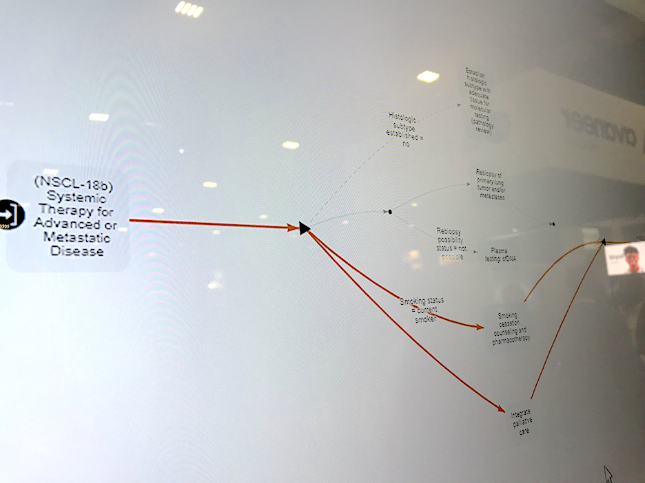
Siemens' Digital AI Pathway Companion for lung cancer uses AI to pull in all the relevant data and prior exams for a patient and offers graphics for the direction care should go based on current guidelines.
Deeper Integrations With Third-Party Imaging Analysis Application
It is not just AI, PACS and CVIS vendors are also looking at adding more deep integrations with third-party vendor applications. They say they want to move away from any interfaces that require opening a separate software, extra login, or workstation. This also will make everything seamless to improve workflow efficiency and the flow of data in the reporting system, archive and EMR access.
A good example of this at HIMSS was Siemens Healthcare showing a new, deep integration with echocardiography strain imaging analysis provider Epsilon Imaging. The integration eliminates the need for a separate login and the Epsilon software is now just a tool option available in the syngo system. The software automatically quantifies the wall motion strain in the right and left ventricles and the quantification numbers are carried over into the Siemens cardiac ultrasound report.
Strain can be used to assess cardiac function more precisely than regular cardiac ultrasound exams. It has seen wide use in cardio-oncology programs, assessing a baseline cardiac function and performing serial exams over the course of a cancer patient’s chemotherapy regimen to catch cardio toxicity issues early, to avoid having the patient go into heart failure.
Reimbursement was recently added for strain in the U.S., which has caused rapid adoption of the technology. Epsilon is seen as the key vendor in this space, so Siemens and Fujifilm are now working with the company for closer integrations
Cerner highlighted a deep integration with third-party vendor Quera in the in the interoperability theater area at HIMSS. The partners connected data from several disparate systems in a healthcare system to create a single view of a heart transplant patient's data for case review and ongoing care of that patient. The companies said transplant patients often no have data that is needed for a complete review of their case and care spread across several disparate health IT systems. This software integration pulls all the parts needed from these various systems into one viewer.
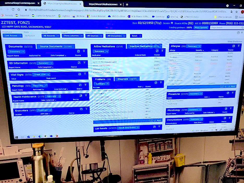
Cerner highlighted a deep integration with third-party vendor Quera in the in the interoperability theater area at HIMSS. The partners connected data from several disparate systems in a healthcare system to create a single view of a heart transplant patient's data for case review and ongoing care of that patient.
Related HIMSS Health IT Content:
VIDEO: Cardiology AI Aggregates Patient Data and Enables Interactive Risk Assessments
VIDEO: Examples of Improved PACS Workflow to Aid Speed and Efficiency
Photo Gallery of New Technologies at HIMSS 2021
VIDEO: Importance of Body Part Labeling in Enterprise Imaging — Interview with Alex Towbin, M.D.
HIMSS 2021 Showed What to Expect From In-person Healthcare Conferences During the COVID Pandemic
VIDEO: Coordinating Followup for Radiology Incidental Findings — Interview with David Danhauer, M.D.

 March 03, 2026