OCT has limitations where IVUS can still offer superior imaging.
There are many advantages to using optical coherence tomography (OCT) intravascular imaging. However, interventionalists should not rush to trade in their intravascular ultrasound (IVUS) systems, said Michael R. Jones, M.D., FACC, director of the Central Baptist Hospital Heart and Vascular Institute, Lexington, Ky.
Jones is a fan of using intravascular imaging in PCI cases and has used IVUS for about 15 years. He transitioned to OCT when his facility purchased its first system in August 2010, and now uses it for the majority of his cases.
“This is a new technology to look at structures in the vasculature and offers very accurate measurements,” Jones explained. “Both IVUS and OCT allow us to look in living, beating hearts. Both of these techniques are used to make measurements for lesion length and lumen size, but OCT is being shown in studies to be more accurate.”
OCT was developed by Massachusetts Institute of Technology (MIT) 20 years ago as a way to examine the retina in ophthalmology. Jones said the technology also has made a big impact in the art world as a way to detect forged paintings and to examine images sketched or painted under the surface layer of paint.
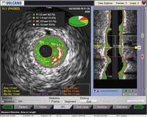
With intravascular imaging, Jones said OCT offers clear, photographic quality images, as opposed to grainy, lower-resolution IVUS images. He said many interventionalists are “confounded” by the speckled IVUS images, which are sometimes difficult to interpret. Fine details, such as stent strut apposition and thrombus, can be difficult to detect.
“The two technologies are analogous in that they send out energy waves — OCT uses light and IVUS uses sound waves — into the vessel wall and that energy is sent back to the catheter to reconstruct an image,” Jones said. “The wavelength of light is much shorter and much faster than sound waves.” For this reason, he said OCT has a resolution 10 times greater than IVUS (OCT 10 microns, IVUS 100 microns).
“There is a real ‘wow’ factor when interventionalists first see the OCT images,” Jones said. “The analogy is going to the lake to go fishing and you drop your fish finder in the water, which is sound wave-based like IVUS, and then you try to figure out from the blips what are fish and what are rocks. But OCT is equivalent to dropping a high-definition color TV camera in the water instead. You can just see better and you can see things like thrombus and thin-cap athromas.”
Being able to visualize thrombus and vulnerable plaque may change the nature of how patients are treated, Jones said.
Watch the video interview "Advances in Intravascular Imaging at TCT 2015"
Read an updated 2015 article about OCT and IVUS technology advances
Workflow, Speed and Function
Jones said OCT is easier and faster to set up and use than IVUS. The St. Jude Medical C7 OCT console is lighter and smaller than current IVUS consoles and it boots up in 30-40 seconds. The system’s Dragonfly imaging catheter also has a smaller size advantage at 2.7 French, whereas Volcano’s Eagle Eye IVUS catheter is 3.5 French and Boston’s IVUS catheter is 3.2 French.
OCT uses a contrast flush to clear the blood column and allows a clear path for the light beams to emit, reflect and be detected. Unlike IVUS, OCT is obstructed by blood. The system uses an automatic pullback at a speed of 20 mm per second, with a pullback length of about 50 mm. The automation and speed is necessary to follow the contrast injection. In comparison, the speed of an automated IVUS pullback is about .5 to 1 mm per second.
The higher OCT resolution makes it easier to evaluate stent strut apposition against the vessel wall. It also allows clear imaging of neointimal stent strut coverage in follow-up exams and allows much better edge detection than IVUS, Jones said.
“I think it has the potential to replace IVUS because it has better resolution,” Jones said.
Issues With OCT
Despite his very enthusiastic endorsement of OCT, Jones said there are some drawbacks. OCT requires additional contrast use. Saline can be used in combination with contrast, but Jones said contrast just works better at clearing blood than saline alone. Extra contrast may present safety issues in patients with kidney impairment.
“You will have some patients who have renal insufficiency and with these patients you should use IVUS,” Jones said.
An Achilles’ heel of OCT, where the system cannot image well, is aortic ostial lesions. There is no way to clear the blood from the aorta at the entrance to right or left main arteries, so Jones said it is difficult to get clear images of these areas. In these cases, he still relies on IVUS.
“The question I always get from interventionalists is if they can trade in their IVUS machine and just use OCT,” Jones explained. “My answer is you still need your IVUS machine.”
Comparing Tissue Image Penetration
Another advantage of IVUS is its penetration of 4-8 mm inside the vessel wall. The light-based OCT technology can only penetrate about 2-3 mm. If a vessel has significantly remodeled due to plaque burden, the outline of the true lumen disappears on the OCT image and the interventionalist is left to connect the dots in his mind as to the extent of the plaque.
However, Jones said this really does not matter in day-to-day use. “If you need to measure plaque burden from the lumen to the adventitia, you might want to use IVUS. But, we are largely ‘lumenologists,’ so we really are concerned just about the lumen. Our strategy is not changed by the amount of plaque. We don’t try to make the vessel as it was before the disease. We find a reference segment of the vessel without plaque and we size our balloons and stents based on that lumen size.”
Comparing Technical Specifications of OCT vs. IVUS
DAIC created apples-to-apples comparison charts of technical specifications for IVUS and OCT systems. These require a log-in, but it is free and only takes a minute.
The IVUS Systems chart can be accessed at www.dicardiology.com/content/ivus-systems
The IVUS Catheter chart can accessed at www.dicardiology.com/content/ivus-catheters
The OCT Systems chart can be found at www.dicardiology.com/content/oct-systems
The OCT Catheter chart can be accessed at www.dicardiology.com/content/oct-catheters

 June 18, 2026


