Interventional cardiologists know well the phrase “change is the only constant.” This is especially relevant as innovative software solutions continue to emerge which provide value on two impactful fronts — clinical workflow and patient outcomes.
Cardiac magnetic resonance imaging (MRI) software tools assess different structures of the heart, its function and condition, and that of peripheral arteries, all valued in both scientific research and clinical work.
The cardiac imaging software market is expected to reach $704.32 million and register a CAGR of 8.54% during the forecast period of 2021 to 2027, according to a Market Research Future forecast report. The growth is attributed to the increasing prevalence and rising healthcare expenditures for cardiovascular disease and growth in the elderly population. The report noted the advancement of artificial intelligence (AI) creates opportunities in the healthcare sector to obtain more sophisticated information from imaging and to find complex patterns in available data sources. The data gathered supports the diagnosis and treatment of patients with cardiac-related diseases, a leading cause of increasingly costly hospitalization.
Innovative Features
In reviewing the market leaders, one vendor client referred to this technology as “a quantum leap moving forward.” Increasing, physicians are finding that leveraging AI tools supports both better diagnoses and improved workflow. Those in cardiac imaging are experiencing a transformation of care delivery powered by AI.
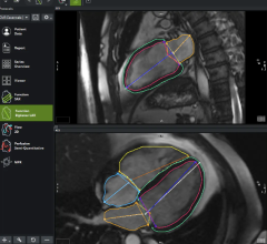
Medis Imaging reports several innovative features have been added to the latest Medis Suite MR. These include the automatic creation of motion corrected T1 Maps providing T1 values from the viewer, a new clinical parameter, and Inward Displacement for regional ventricular dysfunction. The Medis Suite MR now offers a new research parameter, Hemodynamic forces (HDF) for the evaluation of Intra-Ventricular Pressure Gradients, and AI deep learning contours for Left Ventricle in the Long axis, providing GLS in a more reproducible and quicker manner. Enhanced vectors in 4D Flow allow users to visualize the blood flow direction. Other features include a fast and efficient blood flow analysis in 2D and 4D; AI based Left Ventricle and Right Ventricle contour detection for function analysis; and seamless integration in healthcare IT environment and an easy connection with the DICOM network.
Productivity and Customization
Reducing complexity and increasing productivity are key features of the syngo.via tool developed by Siemens Healthineers. The company continues to advance digitalization in healthcare by collaborating with market leaders in reporting and EHR, allowing the software to offer users valuable guideline support. Multi-modality reading, fast 3-D results speed up daily routines, while AI-enabled features support better report creation. Automatic, real-time findings integration and intelligent support assist in generating structured and consistent reports as syngo.via provides actionable image-based results to navigate care delivery. Its schematic overview of data exchange between reading, reporting and EHR provides added value, and simplifies the diagnostic process. Syngo.via seamlessly integrates advanced tools and algorithms, offering a centralized, intelligent, and customizable control center with everything in one place. Its syngo.via OpenApps expands reading capabilities with an open platform.
Workflow Efficiencies
Several workflows can be selected within Caas MR Solutions software package offered by Pie Medical Imaging. Its MR 4D Flow provides analysis of 3D PC-MR images of the heart and major vessels, while the Caas MR 2D Flow features tools to quantify blood flow in the major vessels using 2D PC-MR images. MR images of the heart can be analyzed for functional, viability and perfusion analysis with Caas Functional Analysis and Caas Tissue Characterization. Quantification of strain parameters such as global longitudinal strain (GLS), global circumferential strain (GCS), and global radial strain (GRS) based on the deformation of the myocardial wall is enabled with the Caas MR Strain.
The company’s activities in quantitative analysis software, with Cardiovascular Angiographic Analysis Systems (Caas), started in 1984. In 1995, Pie Medical Imaging was founded, and in 1998, became part of the Esaote group. In 2011, 3mensio Medical Imaging was acquired and Pie Medical Imaging now markets the 3mensio product range next to its Caas products.
Vascular Technology
Bringing together experts in the fields of circulatory fluid dynamics and computer modeling, VasSol was founded on more than a decade of multidisciplinary research and clinical work conducted at University of Illinois at Chicago. Its vascular analysis technology features VasSol’s Non-invasive Optimal Vessel Analysis, NOVA, utilized for the diagnosis and treatment of vascular conditions since its first clinical application in 1997. It incorporates interactive 3-D images for a fully rotatable, 360° view of vasculature that allows for precise identification of each vessel for volumetric blood flow calculation.
Keeping up with the changes in Cardiac MRI software is a critical requirement for clinicians striving to advance workflow, diagnostics and patient care.
View the Cardiovascular MRI Analysis Software comparison chart here

 April 10, 2026