Toshiba America Medical Systems' Aartida
Heart disease is the leading cause of death in Americans, claiming hundreds of thousands of lives yearly. The ability to diagnose and treat patients sooner and with more accuracy is key to winning the war against heart disease. Advancements in ultrasound — one of imaging technology’s most affordable and available modalities — are leading the charge by bringing enhanced diagnostic and treatment accuracy to more patients.
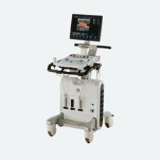
Cardiologist Wolfgang Fehske, M.D., St. Vinzenz Hospital, Cologne, Germany, relies on GE Healthcare’s Vivid S6 cardiovascular ultrasound for applications throughout the hospital.
What makes the Vivid S6 so attractive, he says, is it is a small, mobile scanner packed with high-end technology. Although specifically designed as a dedicated cardiovascular solution, it can be used at various locations beyond the cath lab, including the ICU, the EP lab, the OR and for ultrasound-guided procedures at the bedside.
“The Vivid S6 is also completely compatible with the advanced network technology of the GE EchoPAC system, which is the benchmark in digital imaging,” said Dr. Fehske. “It is important to be able to review all prior data on a patient no matter where you are.”
When the hospital used exclusively larger ultrasound units, they performed fewer exams in the ICU and the cath lab. In Dr. Fehske’s experience, Vivid S6’s mobility and networking compatibility are strong arguments for better patient care.
For physicians requiring enhanced understanding of real-time, 4D anatomy and function, GE offers the Vivid 7 Dimension complete with LV quantification tools, including 4D LV volume on-board. End-users report the system’s features “support an intuitive impression of morphologic anatomic structures in a 3D image and therefore simplify interpretation and 3D navigation impressively.”
Having worked with ultrasound for nearly 30 years, Dr. Ernesto Salcedo, M.D., associate professor of medicine and director, echocardiography laboratory, University of Colorado Hospital, Denver, has witnessed firsthand the technology’s evolving capabilities. The hospital has been using 3D real-time (3D/4D) TEE echocardiography, the Philips’ iE33 system, since May 2007. In the OR, Dr. Salcedo believes the technology’s greatest potential is for surgical mitral valve repair. Standard therapy for treating severely impaired heart valves used to involve implanting an artificial valve; now the goal is valve repair versus valve replacement, he says. But, you must be able to determine which valve is repairable. 3D/4D TEE gives surgeons views of the heart that aren’t possible with two-dimensional data.
“Surgeons can now determine - before surgery - which valve is affected and in what area, which in turn, helps them determine the best course of treatment. Intraoperatively, the technology allows the surgeon to immediately see the results of the repair, and provides good information on the evolution of ventricular function,” Dr. Salcedo said.
Ultrasound vs. CT, MR
How do the newer features of cardiac ultrasound stack up against the benefits of other imaging modalities?
“Ultrasound is the best modality for frontline, immediate return on diagnostic information,” said John Davidson, senior director, cardiology, ultrasound division, Siemens Medical Solutions.
“Today’s cardiac ultrasound technology offers the benefits of immediate detection and reporting, being able to look at regional wall motion abnormalities in real time and assessment of valve structure and morphology,” Davidson said. Other imaging modalities attempt to acquire these end points but they accomplish them over a period of time and they require the patient to travel to the source of the imaging, whereas ultrasound is transportable to the point of care. As a result, the workflow, costs and turnaround times are much more favorable with ultrasound, he says.
“CT is great for analyzing the coronary arteries and for perfusion, but can’t provide a real-time assessment of heart function. With some scanners you can’t see the entire heart and so you have to stitch it together or use some other type of post-processing technique in order to see what’s going on,” added Gordon Parhar, director of cardiac ultrasound with Toshiba America Medical Systems.
“While there is a lot of buzz in the market about CTA, ultrasound is here to stay,” he said. “It’s faster and more cost effective than CT and provides physiological images and data — things that CT cannot, and it does so without any radiation.”
MR tagging has long been considered the gold standard for detecting wall motion abnormalities. But the technology is very expensive, has gating problems and is not always tolerated well by patients. With John Hopkins University, Toshiba is trying to validate strain measured by ultrasound against strain measured by MR.
“Echocardiographic wall motion assessment is still largely subjective. Toshiba’s goal is to find a quantitative way of determining wall motion abnormalities that may lead to earlier diagnoses,” said Parhar.
He said they developed a wall motion tracking technique that is not subject to angle-dependent Doppler limitations and can detect and quantify wall motion defects that are too subtle to be picked up by the naked eye.
Getting the “Full” Picture
“Cardiology is all about temporal resolution,” said Davidson. “Ultrasound has prospered in the imaging world because it provides immediate return on diagnostic information in a real-time mode.”
He said Siemens is developing an architectural capability to enable acquisition of 90-by-90 degree pyramids at 20 volumes/second, every second; in other words, full-volume, true volumetric imaging in real time. This capability is expected to greatly enhance current 3D/4D ultrasound imaging, because it eliminates the need for ECG gating.


 May 19, 2026