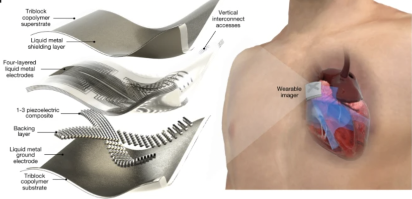
Exploded view of the cardiac ultrasound imager.
January 30, 2023 — Heart disease is the leading cause of death worldwide. In the U.S., it is estimated that someone dies every 34 seconds from cardiovascular disease. Diagnosis for heart disease remains a challenge, as most methods only provide a snapshot of cardiac function, which may not be representative of overall heart health. Improvements in diagnostic technologies could help catch heart disease in earlier stages, potentially preventing numerous deaths.
After years of research, an NIH-funded team has developed a wearable cardiac ultrasound imager that can non-invasively capture real-time images of the human heart for an extended period of time. The patch, which is about the size of a postage stamp, has comparable performance to a commercial ultrasound device. What’s more, the imager can be worn during exercise, providing valuable cardiac information when the heart is under stress. The prototype ultrasound patch was reported today in Nature.
“While existing wearable patches in development can capture things like heart rate and blood pressure, they are not designed to provide in-depth information about heart function,” said Randy King, Ph.D., a program director in the Division of Applied Science & Technology at NIBIB. “This innovative ultrasound device gives critical insight about the heart in real time, providing clinicians with detailed, actionable cardiac information. Combined with its thin design and stretchable properties, this patch could pave the way for continuous, non-invasive cardiac monitoring.”
Traditional cardiac ultrasound imaging requires physical rotation of the transducer, so that specific cross-sections of the heart can be visualized and evaluated by a clinician. This prototype patch, however, has two perpendicular piezoelectric arrays that can be independently controlled. This allows a clinician to image multiple standard views of the heart without any intervention or repositioning of the patch. Liquid metal electrodes connect the tiny arrays so that each element can be individually manipulated, and a type of silicone fills the interior of the device, which makes the entire patch highly stretchable and mitigates the need for an ultrasound gel. Taken together, these design elements allow the patch to maintain close contact with the skin at all times, overcoming the limitations of rigid ultrasound devices.
The researchers first needed to evaluate how their wearable patch compared with a traditional ultrasound device. Using a human tissue phantom model, they characterized the properties of their patch, such as its spatial resolution and its signal-to-noise ratio, and found that these characteristics were similar to a commercial ultrasound device. Next, they used their patch to image the heart of a human subject. Looking at four standard views of cardiac anatomy, they found that the images generated by their patch were similar to those generated by the commercial ultrasound device.
After characterizing the performance of their patch, the researchers evaluated its utility during exercise—specifically, how it fares during a stress test. Traditional stress echocardiography evaluates images of the heart before and after intensive exercise (as holding an ultrasound probe over the chest by hand and maintaining a stable position is impossible during exercise). The patch was used to monitor the cardiac performance of one healthy participant during a rigorous exercise session. The researchers found that the patch could capture the activities of the left ventricle (the heart chamber that pumps oxygenated blood throughout the body) without any interruption.
“Our patch allows us to evaluate heart performance throughout exercise, providing valuable information about the heart when it is under high stress,” explained senior study author Sheng Xu, Ph.D., an associate professor at the University of California San Diego (UCSD). “This could allow for real-time visualization of heart anomalies as they manifest, which might be missed under normal stress test conditions,” he added. Further, the participant wore the patch for 24 hours without any allergy or skin irritation, illustrating its utility for day-long wear.
Finally, the researchers wanted to determine if their patch could be used to calculate key cardiac functions, such as stroke volume, cardiac output, and ejection fraction (which are all related to how much blood is pumped out of the heart and how efficiently the heart is working). Using deep learning, they extracted specific features from ultrasound images taken with their patch and trained a model to reliably extrapolate these cardiac metrics. “The ability to non-invasively and continuously monitor such cardiac functions over a 24-hour period could revolutionize the field,” Ray Wu, a graduate student in the Xu lab and coauthor of this study, said.
An important next step in the development of this patch is to make it entirely wireless. “As of now, our patch requires cables to supply it with power, control the piezoelectric transducers, and transmit the ultrasound data to a back-end system,” explained first study author Hongjie Hu, Ph.D., a postdoctoral researcher at UCSD. “Our immediate goals are to construct a built-in power source for our patch and to develop a system for wireless communication between the patch and a remote device, such as a central workstation,” he said.
“While this wearable ultrasound patch may be in the early stages, its potential to non-invasively provide comprehensive, real-time cardiac information could transform health care in the future,” said Xu.
For more information: https://www.nibib.nih.gov/

 May 13, 2026