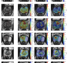
Periannular uptake. Representative examples show hybrid, fused PET/MRI in patients with findings suggestive of periannular uptake. (A) Axial view with uptake in basal lateral and basal septal regions, surrounding mitral annulus. (B) T2 values are globally increased, especially in basal septum. (C) Axial view with increased 18F-FDG uptake in regions surrounding mitral annulus. (D) T1 values are increased in basal septum. (E) Sagittal view with increased uptake in basal segments surrounding mitral valve annulus. (F) ECV values are increased, especially in basal to mid-septum. SUV scale is 0–5 g/mL. AHA = American Heart Association; ECV = extracellular volume.
July 21, 2025 — Long COVID patients with abnormal cardiopulmonary PET/MR findings may be more likely to develop heart and lung diseases, according to new research published in the July issue of The Journal of Nuclear Medicine. The study — which included the largest cohort of long COVID subjects with cardiopulmonary symptoms with the longest follow-up duration — suggests that abnormal cardiopulmonary PET/MR should be considered a risk factor for future cardiac and pulmonary disease and that closer monitoring is warranted in these patients.
To date, the COVID pandemic has seen more than 400 million people infected worldwide, with more than 80 million infections and approximately one million deaths in the United States. Long COVID has emerged as a major public health challenge, affecting between one-third and two-thirds of patients many months after recovery from the initial COVID infection.
“Unfortunately, the long-term consequences of long COVID remain unknown,” said Maria Giovanna Trivieri, MD, PhD, FACC, FRCPC, associate professor of medicine (cardiology) and associate professor of radiology at the Icahn School of Medicine, Cardiovascular Research Institute, Biomedical and Molecular Imaging Institute, in New York, New York. “In this study we sought to use advanced cardiopulmonary imaging to assess for evidence of cardiac and lung abnormalities, vascular injury, and inflammation in patients with long COVID.”
The study included 98 patients with a history of COVID infection, persistent cardiopulmonary symptoms nine to 12 months after initial infection, and a clinical assessment compatible with long COVID. A control group that included subjects with a history of severe COVID but without cardiopulmonary symptoms at recruitment was also characterized. Both groups underwent cardiopulmonary 18F-FDG PET/MRI and dual-energy CT (DECT) of the lungs. A plasma protein analysis was also conducted for a subgroup of patients.
PET/MRI was abnormal for 57 percent of subjects, showing myocardial, pericardial, periannular, and vascular uptake, none of which was present on the PET/MR scans of the control group. Ninety percent of subjects presented abnormalities in DECT, including pulmonary infiltrates and abnormal perfusion. Analysis of plasma protein concentrations showed significant differences between the long COVID and the control groups, and the plasma protein profile was significantly different among long COVID patients with abnormal and normal PET/MR scans. During the four-year follow-up period, PET/MR abnormalities were more frequent in patients that subsequently developed heart failure, mitral regurgitation and pulmonary hypertension.
“The results of the study should raise awareness in the clinicians to elicit a proper history that includes prior COVID infection and long COVID symptoms,” stated Ana Devesa, MD, PhD, former postdoctoral fellow at Biomedical and Molecular Imaging Institute, in New York, and group leader at Centro Nacional de Investigaciones Cardiovasculares (CNIC) in Madrid, Spain. “If a temporal link is identified between symptoms and timing of infection, further evaluation might be appropriate. ”
For more information, visit www.snmmi.org.

 April 27, 2026