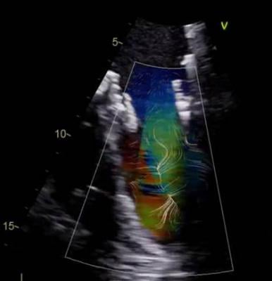
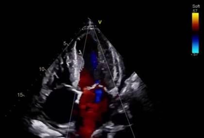
GE’s blood speckle imaging (BSI) software offers the ability to view a graphical representation of the blood cells’ trajectories.
Cardiologists are constantly on the lookout for new methods to examine the heart using new imaging technology advances. Many of the limitations in echocardiography has been due to the limits of image processing speeds. This has limited the image quality based on how much data can be processed and rendered on the ultrasound system screen in real time.
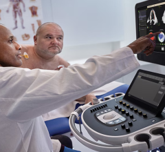
Two years ago, Bijoy Khandheria, M.D., medical director of the echocardiography laboratory at Aurora St. Luke’s Medical Center in Milwaukee, Wis., and his team were introduced to a new echocardiography software beamformer image reconstruction platform from GE Healthcare. The cSound technology allows imaging processing that equals the same amount of data equivalent to playing an entire DVD in just one second, in real-time.
Recently, Khandheria was equipped with the new Productivity Elevated release on GE Healthcare’s Vivid E95. “This is a true leap forward from what was already an excellent cardiac ultrasound imaging platform,” Khandheria explained.
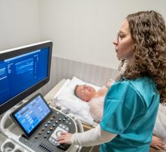
On any given day, more than 100 patients are scanned in St. Luke’s echo lab, each with a different set of medical issues. The ability to properly assess, diagnose and treat these patients. Khandheria said using this new system has enhanced the lab’s productivity and efficiency, and it helps improves patient care overall.
In one recent case, Khandheria saw a patient who had been told his heart activity looked normal for the last three years. But with this new system, Khandheria found irregular activity in the left ventricular that eventually led to a diagnosis of non-compaction – a rare genetic disease of the heart. By catching this early, the team altered treatment and closely monitored the patient.
“We’ve seen several potential benefits to the new workflow features on this ultrasound system, including software enhancements and automation that make it easier for the sonographer to acquire the images and even speed up the process without losing image quality,” noted Khandheria. “In fact, we are able to save three to five minutes per study. When performing several studies every day, this adds up to a significant amount of time.”
These cSound enabled automated features help streamline both routine and advanced studies. The time saved can be used to evaluate critical cases and deliver high-quality, efficient care.
A recent advanced visualization introduction for the system in 2017 is GE’s version of vector flow imaging, which allows cardiologists to clearly see how blood flows through the heart. This is opening up new research into how disrupted blood flow may lead to or accelerate some diseases, including valvular regurgitation, formation of atherosclerotic plaques and heart failure. GE’s blood speckle imaging (BSI) software offers the ability to view a graphical representation of the blood cells’ trajectories. This new way to image patients’ hearts excites Khandheria the most is the ability to view a graphical representation of the blood cells’ trajectories with blood speckle imaging (BSI).
“This is the next true breakthrough, improving efficiency and, ultimately, patient care,” Khandheria said. “It opens a whole new vista of learning what is happening in the heart and cardiovascular physiology.”
Read more about these and other advances in cardiac ultrsound technology at the 2017 American Society of Echocardiography (ASE) conference in the blog "A Glimpse Into the Future of Cardiac Ultrasound."
Watch the VIDEO "Editor's Choice of Most Innovative New Cardiac Ultrasound Technology at ASE 2017."
Read the related article "Reducing Cardiac Ultrasound Exam Times."

 June 15, 2026