October 2, 2009 – Cardiovascular magnetic resonance (CMR) in the evaluation of patients with heart failure (HF) “will expand substantially in the coming years,” according to the authors of a study published in October 2009 issue of the Journal of American College of Cardiology.
In the study, The Role of Cardiovascular Magnetic Resonance Imaging in Heart Failure, the authors emphasize the efficacy of CMR in assessing myocardial anatomy, regional and global function, and viability, and assessment of perfusion and acute tissue injury, and notes that CMR often “reveals the underlying etiology of heart failure, and its high measurement accuracy makes it an ideal technique for monitoring disease progression and the effects of treatment.” They add that increasing evidence that “the prognostic value of CMR-derived parameters in heart failure is rapidly emerging.”
“We predict that most patients with HF will eventually undergo CMR imaging as part of the diagnostic workup and to guide management and stratify risk,” the authors wrote.
The researchers listed the proposed indications for the use of CMR in HF patients that are likely to further expand in the near future. These indications include:
- Serial assessment of biventricular structure, size, and function (anatomy, LV/RV volumes, global and regional systolic function, mass)
- Evaluation of native and prosthetic cardiac valves (planimetry of stenotic disease, estimation of peak stenotic velocities and gradients, quantification of regurgitant disease)
- Evaluation of cardiac masses, differentiation between tumor and thrombus
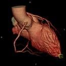
- Evaluation of great vessels and pulmonary veins
- Determination of the location and extent of acute (including no-reflow regions) and chronic myocardial necrosis
Viability assessment before revascularization (LGE or low-dose dobutamine)
- Determination of the area at risk in patients with acute myocardial infarction and the salvageable area post-revascularization with percutaneous coronary intervention
- Identification of the presence and quantification of the extent of inducible ischemia (vasodilator perfusion or high-dose dobutamine stress CMR)
- Evaluation of suspected anomalous coronary origins (MR coronary angiography)
- Differentiation of ischemic versus nonischemic cardiomyopathy
- Evaluation of myocarditis
- Evaluation of specific cardiomyopathies (in vivo tissue characterization)
- Assessment of mechanical dyssynchrony before resynchronization therapy
- Patients with technically limited images from echocardiogram
- Discordant information that is clinically significant from prior tests
Plus, in their conclusion, the researchers anticipate that “improvement in software and hardware will further shorten scan times and allow the use of real-time imaging with better spatial and temporal resolution. New CMR techniques aiming at the identification and quantification of diffuse fibrosis will further improve the in vivo assessment of myocardial pathology.”
They pointed to other growth for CMR that include: novel CMR contrast agents, stem cell therapy monitored by CMR, interventional CMR to guide therapeutic procedures, and finally MR spectroscopy to assess metabolic changes in the failing heart and monitoring of the response to novel forms of metabolic therapy.
Reference: Theodoros D. Karamitsos, M.D., Ph.D.,* Jane M. Francis, DCC(R), DNM,* Saul Myerson, M.D.,* Joseph B. Selvanayagam, MBBS, DPHIL,† Stefan Neubauer, M.D.,* Oxford, United Kingdom; and Adelaide, Australia. The Role of Cardiovascular Magnetic Resonance Imaging in Heart Failure. J Am Coll Cardiol, 2009; 54:1407-1424, doi:10.1016/j.jacc.2009.04.094
For more information: www.ocmr.ox.ac.uk

 May 14, 2026