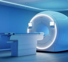
New findings suggest that MRI machines (such as the one pictured above) may help quickly screen stroke patients for acute treatment. Image courtesy of the National Institutes of Health.
May 22, 2015 — According to a new study, small changes in quality improvement procedures enabled clinicians to use magnetic resonance imaging (MRI) scans to diagnose stroke patients before giving acute treatment, within 60 minutes of arrival. MRI scans provide detailed images but take longer to complete than computed tomography (CT) scans, which are commonly used in most centers.
The study, Screening with MRI for Accurate and Rapid stroke Treatment (SMART), was published in Neurology and was supported in part by the National Institutes of Health’s (NIH) National Institute of Neurological Disorders and Stroke (NINDS).
“By making small changes to our processes, we were able to scan suspected stroke patients with MRI and appropriately treat patients within a goal time of 60 minutes. This is an important finding for hospitals, healthcare providers and the public,” said Amie Hsia, M.D., medical director of the Comprehensive Stroke Center at MedStar Washington Hospital Center, Washington D.C., and senior author of the study. “Not only does MRI provide more precise and complete information than the traditionally used CT scan, now we’ve also demonstrated that it is feasible to use from a time perspective.”
National guidelines suggest that stroke patients should receive treatment within 60 minutes of arriving at the hospital. The majority of hospitals rely on rapid CT scans to determine if an individual is eligible for intravenous tPA, the only U.S. Food and Drug Administration (FDA)-approved treatment for ischemic strokes, those caused by blood clots in the brain. If a CT scan shows the patient is having a bleeding, or hemorrhagic, type of stroke, tPA cannot be used as treatment. For many years CT scans were the only imaging tool available in most hospitals, but now MRIs are becoming more widely available.
Clinicians at MedStar Washington Hospital Center and Suburban Hospital, Bethesda, Maryland, routinely work with physician-scientists from NIH and have access to their cutting-edge medical protocols and technologies. The two hospitals use MRI instead of CT scans to screen stroke patients. Although MRI scans can take up to 15 minutes longer than CT scans, they provide clinicians with more detailed information about what is happening in a patient’s brain. Using MRI, clinicians can see early changes taking place during the stroke. In this way, they can see what tissue is at risk and identify blocked blood vessels or subtle bleeding that cannot be picked up by CT.
To reduce the door-to-treatment time, multidisciplinary teams at both hospitals carefully examined the existing processes to identify time-consuming bottlenecks or duplicative methods. By using “lean process interventions,” they found a number of steps that could be eliminated or changed. For example, at MedStar Washington Hospital Center, a lengthy MRI screening form was simplified to three questions; at Suburban Hospital, tPA was put into the medication cart in the MRI suite so that it could be given immediately to patients after scanning instead of returning them to the Emergency Department for treatment.
Once the changes were implemented, Hsia’s team examined whether they had an impact on treatment times for patients. The results indicated that door-to-treatment time was reduced from 93 to 55 minutes, a difference of 40 percent. Over a two-year period, the percentage of patients treated within 60 minutes increased from 13 to 61.5 percent. Further analysis revealed that these changes were due to faster MRI start times.
“There was no difference in the patient characteristics. It was clear the improvements were due to the changes we made in the processes at these two hospitals,” said Hsia.
“A number of the changes that Dr. Hsia’s team assessed were not specific for MRI scans, but were related to general procedures of getting patients ready for imaging as quickly as possible. This suggests that these findings are relevant even in hospitals that do not have emergency access to MRI scanners,” said Walter Koroshetz, M.D., acting director of NINDS. “We will persist in evaluating best practices for acute stroke care to ensure that the greatest number of patients receive treatment as early as possible following stroke.”
Hsia and her colleagues will continue to monitor the door-to-treatment times to ensure they are sustainable. In addition, they plan to continue to evaluate best practices for acute stroke care and look for other improvements to further decrease door-to-treatment times for patients.
This work was supported by the NINDS.
For more information: www.nih.gov

 May 14, 2026