October 18, 2019 — An algorithm developed by faculty at The University of Texas Health Science Center at Houston (UTHealth) can help physicians outside of major stroke treatment centers assess whether a patient suffering from ischemic stroke would benefit from an endovascular procedure to remove a clot blocking an artery.
Results of their clinical study using the algorithm were published online in the journal Stroke.[1]
Endovascular thrombectomy is a procedure that involves threading a catheter through the femoral artery in the leg all the way to the brain, where the clot can be removed mechanically. Since 2015, studies have shown it can improve outcomes for stroke patients, but only if the amount of brain tissue that has been injured is minimal at the time of treatment.
Unfortunately, advanced neuroimaging to detect within hours whether a patient is a candidate for the treatment is limited to emergent magnetic resonance imaging (MRI) or computed tomography (CT) perfusion. That kind of technology and expertise are not available at most community hospitals and primary stroke centers.
“With endovascular thrombectomy, we now have a treatment for ischemic stroke that is really revolutionary. It allows us to take stroke patients from severe disability and return them to an almost normal life,” said Sunil A. Sheth, M.D., corresponding author and assistant professor of neurology with McGovern Medical School at UTHealth. “Unfortunately, the advanced imaging techniques used currently to identify which patients benefit from this procedure are not widely available outside of large referral hospitals. As a result, most stroke patients don’t have access to guideline-based screening for these treatments.”
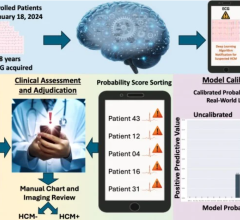
In response, Sheth and Luca Giancardo, Ph.D., senior author and assistant professor at UTHealth School of Biomedical Informatics, developed a machine learning tool that could be used with a widely available imaging technique, CT angiogram. The tool can analyze images by automatically “learning” subtle image patterns that can be used as a proxy for other more advanced, but not readily available, imaging modalities such as CT perfusion. The machine learning architecture, called DeepSymNet, was developed at UTHealth.
To test the tool, the research team identified patients in their stroke registry who had suffered a stroke or had conditions that mimicked stroke.
Of the 224 who had stroke, 179 had cerebral blood vessels that were blocked. The DeepSymNet algorithm learned to identify these blockages from the CT angiogram images, and trained the software to use those same images to define the area of brain that had died, using concurrent acquired CT perfusion scans as the “gold standard.”
“The advantage is you don’t have to be at an academic health center or a tertiary care hospital to determine whether this treatment would benefit the patient. And best of all, CT angiogram is already widely used for patients with stroke,” Sheth said.
For more information: www.ahajournals.org/journal/stroke
Reference
1. Sheth S.A., Lopez-Rivera V., Barman A., et al. Machine Learning–Enabled Automated Determination of Acute Ischemic Core From Computed Tomography Angiography. Stroke, published online Sept. 24, 2019. https://doi.org/10.1161/STROKEAHA.119.026189

 May 20, 2026