January 28, 2016 — A new imaging technique could reduce the need for amputation in patients with critical limb ischemia (CLI), according to a study published in the scientific journal JACC.
British Heart Foundation (BHF)-funded researchers at St. Thomas’ Hospital, London, have developed a new way of mapping blood delivered to the leg muscle immediately after operations on people with severely reduced blood flow to their limbs. Currently surgeons may need to wait days or weeks to see how successful the surgery has been.
They hope that the new technique will ultimately reduce the need for amputations, which can be needed if blood flow cannot be restored.
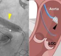
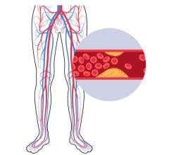
This surgery is used to treat CLI — a serious condition which occurs when the arteries in the limbs become blocked, dramatically reducing blood flow to the feet and legs. Up to 30 percent of patients with CLI eventually have to have the affected limb amputated.
In order to treat CLI, the blocked part of the artery has to be either bypassed during surgery or widened using a stent.
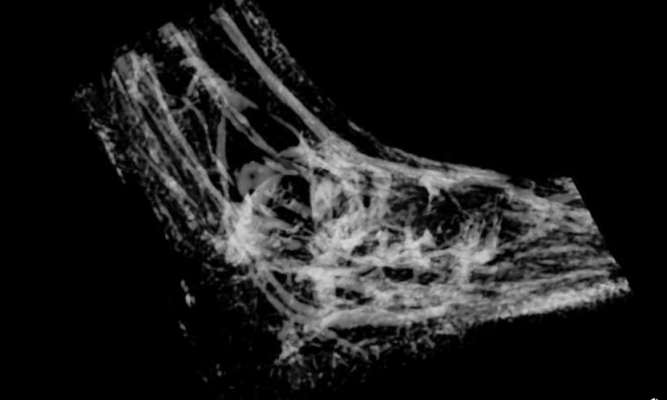
Magnetic resonance imaging (MRI) or computed tomography (CT) scans are used to image the main arteries in the affected limb so a surgeon can understand which area of the artery needs to be operated upon. However, these scans are not perfect — they cannot give the surgeon information about the smallest blood vessels in the legs, or be used to tell how much blood is actually being sent to the limb’s muscle.
During the study researchers used the new MRI-based mapping technique to determine how much blood was reaching the muscles in the legs of 34 people with CLI, before and after treatment, and in 22 healthy people. They then obtained a small amount of leg muscle using a biopsy, in order to check that the results provided by the new technique were correct.
Surgeon Bijan Modarai, who led the research, hopes the new technique could eventually be used to immediately see how much blood flow to the limb had been improved. Currently, the only way of determining the success of surgery is to wait for a period of days to see whether the leg improves.
Modarai, BHF intermediate fellow and reader/consultant in vascular surgery at King’s College London/St. Thomas’ Hospital, lead author of the study, said, “Here at Guy’s and St. Thomas’ NHS Foundation Trust we treat over 500 people with critical limb ischemia each year. By using this new imaging technique we hope to be better placed to offer the best possible treatment to people suffering from this disease and therefore reduce the likelihood of limb amputation.”
Valerie Ellis, 70 from Dartford, had surgery in 2015 to treat critical limb ischemia in her right leg. She said, “The pain in my foot was terrible, I couldn’t lie down or get to sleep at night. I saw my GP and was sent to St. Thomas’ where they did some scans and found that a blood vessel in my right leg was clogged. The surgical team there fitted two stents to widen the artery in my leg and improve the blood flow to my foot.
“It’s brilliant to know that there are researchers out there looking into this very painful disease.”
Prof. Jeremy Pearson, associate medical director at the BHF, said, “The strength of this study is that it shows that this new imaging technique could be used widely to accurately show whether blood flow to the small vessels in the leg is improved sufficiently by stenting or bypass surgery. By using this technique surgeons will quickly gain a clear indication of whether treatment has been successful.”
For more information: www.bhf.org.uk

 February 06, 2026