
Image: Konica Minolta
June 29, 2026 — Konica Minolta, Inc. has announced that a research group has developed a simple and rapid technique for quantifying the severity of pulmonary regurgitation (PR), using Konica Minolta's Dynamic Digital Radiography (DDR). The research group was led by Assistant Professor Yuzo Yamasaki of the Department of Radiology, Kyushu University Hospital, Professor Kousei Ishigami of the Department of Clinical Radiology, Graduate School of Medical Sciences, Kyushu University, Associate Professor Kenichiro Yamamura of the Comprehensive Maternity and Perinatal Care Center, Kyushu University Hospital, and Research Assistant Professor Ichiro Sakamoto of the Department of Cardiology, Kyushu University Hospital (at the time of the study).
This technique is expected to reduce the burden and improve the efficiency of post-surgical evaluation for Tetralogy of Fallot (TOF)*. These findings were published in Radiology. Konica Minolta is also participating in the research group.
Background
TOF is the most common cyanotic congenital heart defect, and treatment generally requires cardiac surgery. Today, many patients survive into adulthood after surgery; however, PR is a common post-surgical complication that, if severe, can increase the risk of right‑sided heart failure and arrhythmias. Lifelong monitoring of PR severity, along with timely pulmonary valve replacement, is therefore essential.
Currently, cardiac MRI is the standard practice for quantifying the severity of PR. However, several challenges limit its accessibility: cardiac MRI equipment is typically restricted to specialized facilities, the examination requires radiographers and radiologists with substantial expertise, and the overall cost of the procedure is high. Additionally, MRI cannot be performed for patients with incompatible pacemakers or those with claustrophobia. Echocardiography is another evaluation technique that is simple and widely available; however, image quality is often poor due to post-surgical anatomical changes, and, because the assessment is primarily qualitative, its reliability in determining severity is limited.
Against this background, there is a clear need to develop a simple and quantitative technique that overcomes the limitations of cardiac MRI and echocardiography.
Study Contents and Results
In this study, a novel technique was developed to quantify the severity of PR by extracting and analyzing temporal changes in pixel values over the pulmonary arteries on DDR as waveforms. In a validation cohort of 58 post-surgical patients with TOF and 14 healthy volunteers, the index derived from this technique (Max PV slope) demonstrated a strong correlation (R = 0.87) with the regurgitation fraction measured by cardiac MRI. For detecting severe PR (regurgitation fraction > 30%), the diagnostic performance achieved a sensitivity of 93%, specificity of 94%, detection accuracy of 93%, and an AUC of 0.98.
This technique is expected to function as a simple, low-burden screening tool to help determine whether cardiac MRI is necessary in patients whose PR severity cannot be reliably assessed using echocardiography. It can also serve as an alternative test for patients who are unable to undergo cardiac MRI due to claustrophobia or the presence of a pacemaker.
While the number of patients with adult congenital heart disease continues to increase worldwide, access to specialized cardiac MRI remains limited in rural regions and areas with limited medical resources. Wider adoption of this technique has the potential to help reduce these healthcare disparities.
These findings were published in Radiology, underscoring the usefulness and value of this new DDR‑based technique.
DDR
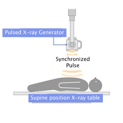
DDR is Konica Minolta’s proprietary X‑ray imaging technology that rapidly acquires a series of sequential radiographs, enabling the motion of organs and other tissues to be visualized as dynamic images. This makes it possible to observe breathing, heartbeat, joint movement, and other physiological cycles in detail, providing more diagnostically useful information than conventional still images. Another major advantage is its ability to capture dynamic information at a radiation dose approximately equivalent to that of two standard chest radiographs (posteroanterior and lateral views).
In addition, DDR is equipped with advanced image-processing and analysis modes that enable functional evaluation beyond simple motion display. These capabilities make it possible to visualize and analyze functional information that cannot be obtained with conventional plain X-ray imaging.
*Tetralogy of Fallot (TOF) is a congenital heart defect characterized by four key features: ventricular septal defect, pulmonary stenosis, over-riding aorta, and right ventricular hypertrophy. It is the most common cyanotic congenital heart defect, affecting approximately 1 in 3,500 newborns.

 June 12, 2026


