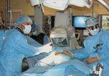
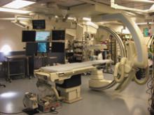
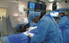
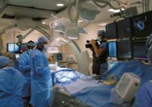
Mortara Instrument offers H-Scribe, a Web-based Holter data transfer from an unlimited number of client recording sites. Data can be transmitted over the Internet or Intranet.
The H3+ digital recorder redefines “small” for Holter recorders while providing 2- and 3-channel recordings, an integrated waveform display, pacemaker detection and up to 48 hours of recording with a single AAA battery. Patients have the freedom to perform their daily activities and even forget they are wearing a Holter recorder, according to the company.
The most recent ELI 350 software introduces the ability to acquire 15-lead ECGs with additional V3R – V4R – V7R, or V7 – V8 – V9, or E1 – E2 – E3 allowing physicians to optimally position the leads according to the patient’s cardiac condition. With the “Best 10” feature enabled, the ELI 350 automatically selects the best 10-seconds in up to 20-minutes of ECG lead connection.
Mortara’s H12+ digital Holter record provides 12-lear data recorded beat by beat on a compact flash memory card for up to 48 hours. The devices features full graphic display to preview ECG waveform during patient hookup.
The Mortara T12S 12-lead ECG digital transmitter integrates pulse oximetry technology that provides continuous transmission of SpO2%, pulse rate and plethysmographic waveform to a receiving patient monitoring system. The T12S is 3 by 4.4 by 1 inch and has a water resistant design.
February 26, 2009 - The first 3D imaging results obtained with a new imaging technology called Magnetic Particle Imaging (MPI) produced reportedly unprecedented real-time images of arterial blood flow and volumetric heart motion, reported Royal Philips Electronics.
February 26, 2009 - In 2006, Americans were exposed to more than seven times as much ionizing radiation from medical procedures as was the case in the early 1980s, according to a new report on population exposure released March 3rd by the National Council on Radiation Protection and Measurements (NCRP) at its annual meeting in Bethesda, MD.
Siemens Healthcare’s Argus 4D VF for 4D visualization of cardiac function. This software enables the visualization and analysis of heart function disorders through the dynamic volume display of the beating heart. With this flexible tool, the cardiac function can be calculated from almost any combination of short-axis and long-axis images.
Siemens Healthcare’s Inline VF (ventricular function) is reportedly the first fully automatic software module for cardiac function analysis with MRI. This module is used to analyze cardiac image data immediately after acquisition and without user interference. Up to now, the image data had to first be transferred to a post-processing computer for manual processing.