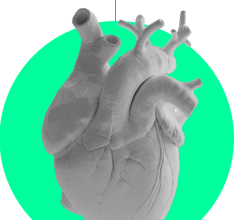
ViosWorks demonstrates the directional flow of blood in the heart and vessels. Color depicts velocity of blood flow.

ViosWorks enables large image datasets of the whole chest to be post processed and evaluated in real-time via cloud technology.
December 15, 2015 — At RSNA 2015, GE Healthcare showcased Viosworks, a new, advanced application to greatly simplify cardiac magnetic resonance imaging (MRI) exams. GE hopes the software will enable cardiac MRI to gain ground in the U.S. market, where it curly only makes up about 1 percent of MRI exams due to the time and complexity.
ViosWorks is designed to help solve several cardiac MR challenges at once. It delivers a three-dimensional spatial and velocity-encoded dataset at every time point during the cardiac cycle, yielding high resolution, time-resolved images of the beating heart and a measure of the speed and direction of blood flow at each location. ViosWorks captures seven dimensions of data (three in space, one in time, three in velocity direction).
It uses a free-breathing scan that can be acquired in less than 10 minutes. A conventional cardiac MRI takes about 70 minutes. ViosWorks can simultaneously provide key elements of a cardiac MR exam: anatomy, function and flow. This provides four advantages.
• The exam is simplified for the patient by using a single 3D free-breathing scan.
• The error-prone and time-consuming aspect of slice positioning is removed.
• Co-registered anatomic images provide the ability to assess cardiac functioning 3D and contextualize
flow abnormalities, and as a result, the cardiac exam can be reduced from over 60 minutes to
approximately 10 minutes or less.
• New algorithms allow large datasets unimaginable before to be evaluated in real-time via the cloud
based Arterys software. Arterys visualization is designed to significantly reduce the amount of time spent on data processing and bring new visualization routines to life.
The software is designed for use on the Signa Pioneer 3.0T, Signa Explorer and Signa Creator 1.5T MRI scanners.
For more information: www.gehealthcare.com

 January 28, 2026
