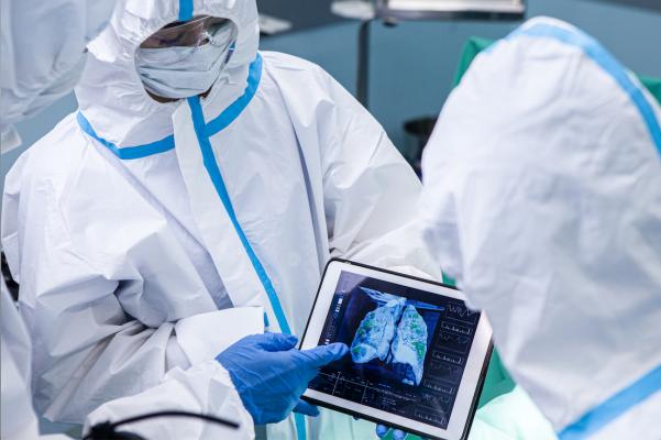
Clinicians reviewing a COVID-19 patient's lung CT that reveals the severity of COVID-caused pneumonia. The impact of COVID on radiology was a major, over arching trend at the 2020 Radiological Society of North America (RSNA) meeting. Getty Images
Not surprisingly, many of the key trends observed at the 2020 Radiological Society of North America (RSNA) meeting focused around COVID-19 (SARS-CoV-2) and the impact it has had on radiology and healthcare. The underlying question throughout the conference was how can the industry take the information from this past year and learn from it?
In addition, new advances in artificial intelligence, MRI automation and simplification, and photon counting computed tomography systems headlined new technologies. Technologies to enable social distancing such as remote operation of scanners, platforms to enable telehealth and remote radiology reading and collaboration also were at the forefront of vendor discussions and booth tours.
Alan Pitt, M.D., a neuroradiologist at Barrow Neurological Institute in Phoenix, Ariz., sees the pandemic as a catalyst for change, and a way to reset how radiologists view healthcare. “We are an enabler of value of public health — taking care of as many people as possible and giving the highest value possible,” he stressed in a Philips Roundtable during the conference. “It’s time to refocus: How many lives can we save? 2021 should be interesting. We need to keep up this level of urgency.”
There were several important takeaways from RSNA20:
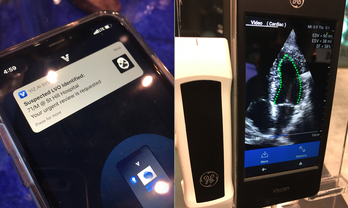
Two examples of FDA cleared AI at RSNA. Left, the Viz.AI software can automatically identify a stroke case as a CT scan is performed and immediately alert the radiologist on call to open a link to look at the case on their mobile device. Right, automated left ventricular ejection fraction using Dia's software on GE Healthcare's vScan point of care ultrasound system. Photos by Dave Fornell at RSNA 2019.
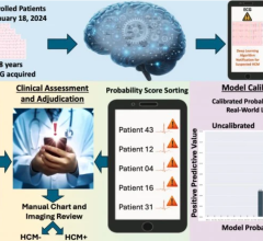
1. A change in attitude and acceptance of AI in medical imaging.
Artificial intelligence allows for more consistent standardization of images. AI systems can help with workflow and boost productivity. At RSNA20, AI was seen integrating into all modalities.
As artificial intelligence is increasingly integrated into real-life workflows, attitudes about AI have changed. At previous RSNA meetings, vendors spent a lot of time explaining how their AI works and how it was validated. Today, this conversation has largely changed to just accepting that an FDA-cleared AI application is legitimate and the radiologists are now asking how it can be smoothly integrated into workflow and what it will do help save them time.
AI is no longer a science project or a 101-level discussion on how the technology can sort pictures of cats and dogs. It has become a real, serious product discussion as these AI applications are integrated into imaging systems and PACS software. Numerous vendors were showing FDA-cleared or 510(k)-pending AI products that can offer a preliminary diagnosis, either to red-flag critical findings and send the exam to the top of a reading list, or serve as a second set of eyes to offer a diagnosis or catch incidental findings.
Several AI algorithms are now FDA-cleared to offer a quick diagnosis of stroke and whether it is ischemic or hemorrhagic. Other FDA-cleared AI on ultrasound systems enables automatic calculation of left ventricular ejection fraction, coaching an inexperienced sonographer on probe placement to get idea echocardiogram views, automation of MRI cardiac quantification, and at software to aid in report completion with auto complete text based on what the AI sees in the imaging.
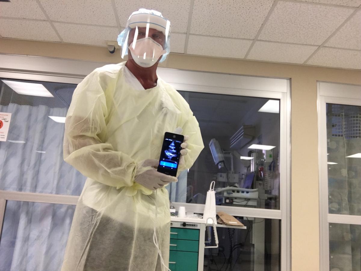
Naples Community Hospital Director of Cardiac Imaging Bill Shirkey showing a point-of-care echocardiogram of a COVID-19 patient he imaged with a GE Healthcare Vscan device bedside in an isolation room. Use of a small handheld device greatly speeds disinfection after the exam and does not require moving a larger cart-based ultrasound system into a room, which may require moving furniture and add exposure time in the room.
2. COVID has caused rapid expansion of point-of-care ultrasound (POCUS).
There was a big question as to the best imaging modality for detection of COVID-19 and later for monitoring critically ill virus patients in the ICU. CT became a front line imaging modality in most emergency departments and mobile digital radiography (DR) X-ray systems saw a big bump is sales in 2020 to enable imaging in patient rooms or dedicated units in COVID wards. But, the imaging modality to see a virtual explosion in 2020 was small, compact point-of-care ultrasound (POCUS) systems.
These included small cart based systems and handhelds. Handheld systems like the Philips Lumify, GE vScan, and Butterfly Network's app-based POCUS that turns a mobile phone into a multi-organ ultrasound system, all saw skyrocket growth. These systems can be placed inside a probe sheath and are much easier to clean after each use than a larger system. Hand-held POCUS systems had a lot of interest prior to COVID-19, but the pandemic showed the extreme value of these tiny imaging systems in a major emergency during the current pandemic.
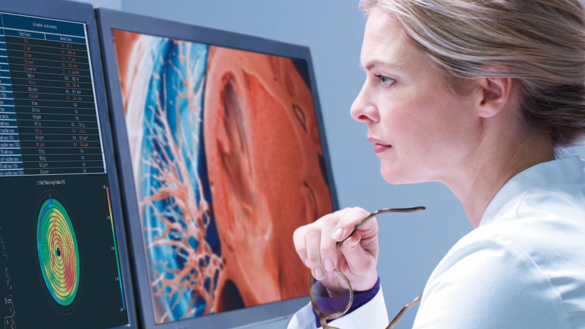
By taking a systems view of imaging, Philips is connecting workflows (patient, acquisition, interpretation, collaboration) across the imaging enterprise to help improve outcomes for patients and enhance the efficiency and work experience of radiologists, cardiologists, technologists and administrators.
3. Telehealth, teleradiology saw explosive growth in 2020.
Radiologists are seeing COVID every day, and it is a lot like Groundhog Day. Pitt says clinicians need to use it as a tool to replan and repurpose. Consistently look at resource allocation. Pitt hopes to see 2020 as a reset on how we view healthcare. “We are an enabler of the value of public health — physicians take care of as many people as possible, and give the highest value possible,” he stated. “We need to refocus — how many lives can we save? 2021 should be interesting. We need to keep up this level of urgency.”
Telemedicine and teleradiology adoption exploded in 2020 due to COVID-19 and it is likely here to stay. This was largely fueled by the Centers for Medicare and Medicaid Service (CMS) creating an emergency rule to allow reimbursement for virtual visits and removing HIPPAA liability concerns so that public commercial platforms like Zoom and Teams could be used. Telehealth likely will permanently alter how healthcare operates, provided CMS grants telemedicine visits permanent reimbursable status.
There was already a large movement before COVID toward enterprise imaging systems to replace traditional PACS or cardiology PACSs. Enterprise imaging systems can be accessed from anywhere over the web and offer the ability to share reports and images across the enterprise for peer review and to referring physicians. These systems are also being used to consolidate several departments' image storage and reporting systems into one central location, which alos aids with integration into electronic medical records (EMRs). The pandemic has increased demand for the remote features and easier remote access to all patient data from internet connect computers at home, remote clinics or in the hospitals.
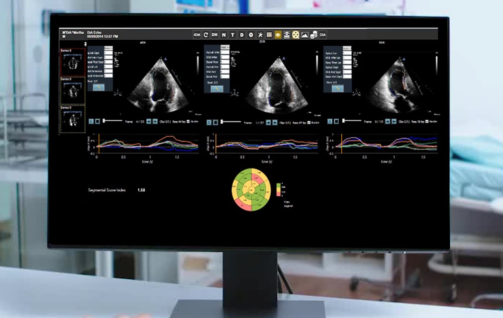
Dia's LVivo artificial intelligence software can help automate many features of echocardiograms to speed workflow and aid novice users. The software is now integrated into the Konica Minolta Exa PACS and is an example of the partnerships between start-up AI vendors and established imaging system vendors.
4. There is a renewed value in partnerships to expand AI in medical imaging.
In 2020, many companies partnered with AI technology vendors to help increase productivity and accuracy. Examples include Canon partnering with Zebra, Glassbeam and Barracuda, and Konica Minolta with 1Qbit and Dia.
Artificial intelligence is a major area of partnering. Large vendors originally hoped to build out a complete menu of AI solutions, but when the reality set in that this was a daunting task that would take decades to complete, there was a rapid switch to partnering with start-up companies that already figured out algorithms, validation and had gained FDA clearance. All of the major vendors are now offering some third-party AI software in its option lineup. Some have even developed an AI marketplace, a sort of AI app store, where they can select which AI they want to try or integrate into their workflow.
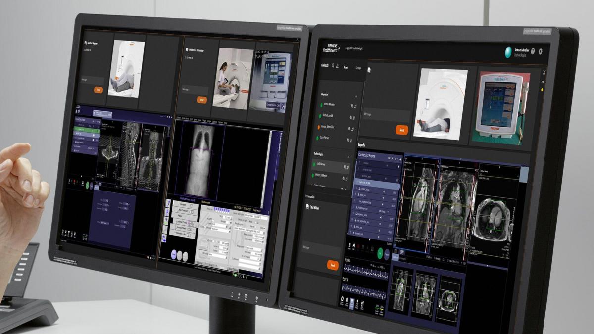
With the Siemens Virtual Cockpit system, RT's can now operate CT and MRI systems without needing to be present in the system's control room.
5. Remote access to imaging scanners driven by COVID.
Several vendors had already developed technology that enabled company technicians to remote into one of their installed imaging systems and help radiology technologists (RT) troubleshoot problems, or to do remote training. Now, in the era of COVID, hospitals are asking if there is any way this technology can be used to allow their RTs can work remotely, operate scanners, or to help other staff members troubleshoot problems, set protocols or take over the scanner operation remotely. This is now being done across some healthcare systems, allowing their own staff members to remotely access scanners, see video of patients on the exam table, regardless if it is the imaging room next door, or at a remote hospital hundreds of miles away.
Siemens modified its Virtual Cockpit system to do just this. RT's can now operate computed tomography (CT) and magnetic resonance imaging (MRI) systems without needing to be present in the system's control room.
Philips Healthcare began the commercial launch of its Radiology Operations Command Center at RSNA 2020, which allows staff to remote into the control rooms of any CT or MRI scanner in their enterprise. It is vendor neutral, so it can interface with multiple vendors' systems. It allows an RT anywhere in the enterprise to enter any of the healthcare systems' MRI or CT control rooms in a few seconds and access cameras to see around the room. They also can remotely take over scanner operations.
Philips said a key use case is if there is a senior technologist who often helps less experienced RTs. Instead of going to a room, or even a separate facility, they can remote in and help the RT through any issues or take control of the system to show them how to do something. The system also records everything for possible later use in training. Another use case is when staff call in sick, especially during the current pandemic where they might be out for up to two weeks. RadNet is now using this system and is currently connected to 24 scanners in the New York City area.
"We have not been able to bring these systems up fast enough," added Vijay Parthasarathy, venture leader for Philips precision diagnosis ventures.
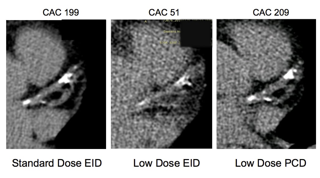
The impact of photo-counting CT detectors on coronary artery calcium (CAC) scoring. Photon-counting detectors will be able to greatly reduce the amount of radiation used to create CT images and will be able to show more detail in those images. This series of images shows CAC scoring changed based on the imaging technology used.
6. Photon counting detectors will be the next major CT innovation.
Several vendors are working on photon counting detectors (PCD) for their next generation CT scanners. The technology has the potential to allow for lower radiation dose while maintaining image quality, or increase the image quality by keeping dose equivalence compared to currently used CT detectors. PCDs also allow the "binning" of photons of different energies, so all CT images created can be post-processed into high or low energy images, or spectral imaging. Some vendors say this detector technology will increase data mining capabilities from multiple-energy detection and contribute to the improved utility and efficacy of AI applications.
While this technology has been researched academically for several years, it appears several vendors are close to commercializing these detectors. At RSNA20, GE Healthcare said photo counting CT detector technology is the way of the future. It purchased the Swedish company Prismatic Sensors AB in November 2020 just prior to RSNA, which is developing photon-counting deep silicon detectors that GE will use on its prototype photon counting CT system. GE hopes to begin operation of this prototype system in the first quarter of 2021.
Samsung is working with a large U.S. university to develop its PCD technology and discussed the new detector at RSNA 2020.
Find out more on this trend in the VIDEO: Photon Counting Detectors Will be the Next Major Advance in CT — interview with Todd Villines, M.D.
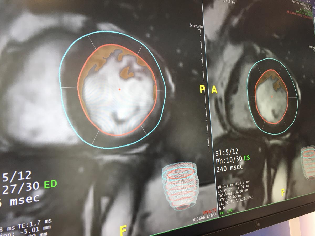
AI automated cardiac MRI image analysis software offered on TeraRecon's Envoy AI platform. Several vendors at RSNA 2020 were showing AI algorithms to automate the contouring of anatomy and quantification, which can greatly speed MRI exams when most of the time consuming work is completed before the radiologist reads the exam. Photo by Dave Fornell at RSNA 2019.
7. MRI may see increased use as it becomes easier to use.
In the past few years at RSNA, MRI vendors have introduced several new technologies to greatly reduce scan times, as well as simplification of MRI scanner operation and protocol setup, greatly reduce or eliminate the need for helium coolant refills and quench pipes, and automation of MRI post-processing. AI is now being introduced for improved image reconstruction to reduce artifact reduction and improve image clarity, and for post-processing automation and quantification. Vendors are banking on these features leading to wider use as these new MRI scanners allow more patients to be scanned per day and their operation and maintenance is greatly simplified.
Leading this trend at RSNA 2020, Siemens introduced its Magnetom Free Max MRI. It is the smallest footprint MRI system to date, and uses only 0.7 liters of helium and does not require a quench pipe, so it can be installed like a CT scanner, rather than previous generations of MRI. It also offers a large 80 cm bore, and is one of the first MRI systems to enable lung imaging.
The vendor is designing this new system to make MRI more affordable, and easier to operate and install, which will open up possibilities for using MRI at urgent care centers or orthopedic offices. Siemens hopes the price point and ease of use may allow centers to buy one of these systems rather than a new or additional CT scanner.
GE, Philips and Canon also introduced AI enhanced MRI image reconstruction technologies.
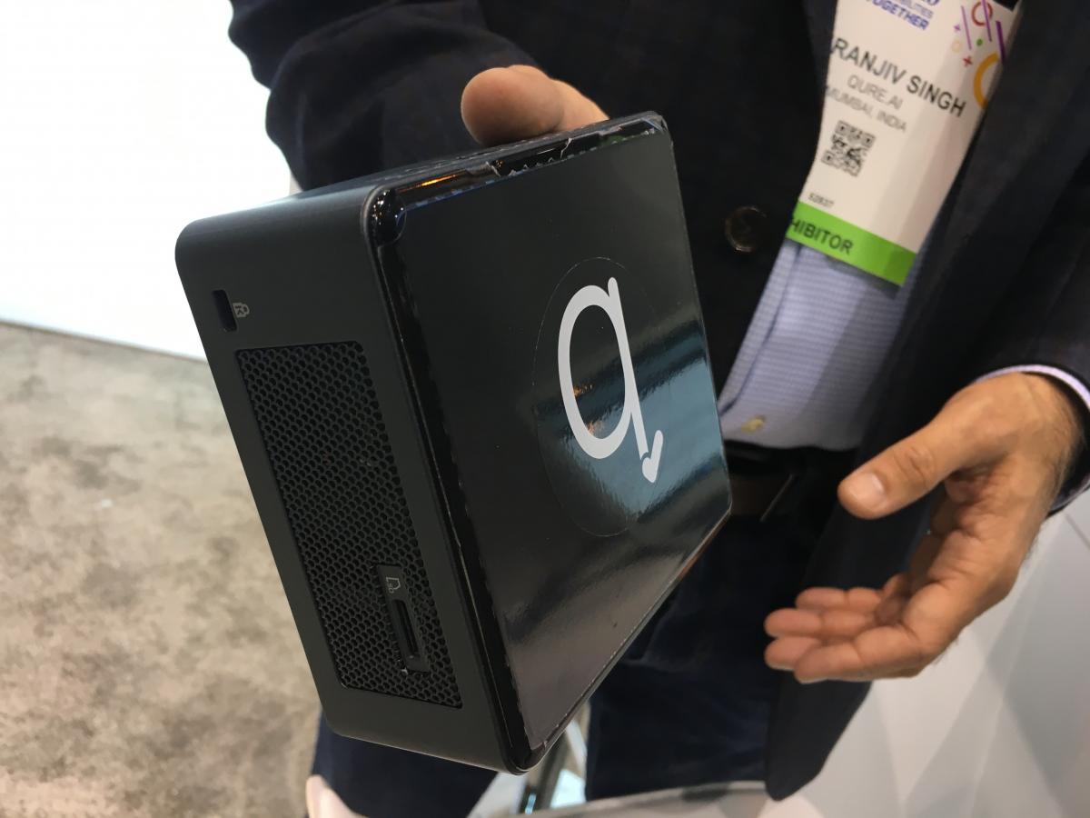
AI vendor Qure.AI has provided its technology to immediately and automatically detect tuberculosis on X-rays to several developing countries with TB detectio programs. However, a big barrier is lack of access to PACS or to the internet in remote areas. So the company created this box that contains everything needed to run its AI algorithm so it can be used in remote areas. This kind of technology is what many vendors say will become a major effort in the coming years to leverage AI to reduce health disparities. Photo by Dave Fornell at RSNA 2019.
8. AI will likely play a major role in next decade to address disparities.
The pandemic past year opened the eyes of radiologists and vendors to wide disparities in healthcare and many areas where innovation is needed. Some sobering statistics were shared during the Philips roundtable regarding the challenges that radiology currently faces:
- Radiology exams with advanced modalities have increased by 43 percent.
- Health data is expected to reach more than 2,000 exabytes in 2020.
- Imaging staff think 23 percent of their work is inefficient and would be better if automated.
- Unnecessary, sub-optimal and repeat imaging cost as much as $12B+ annually.
- There is a growing global shortage of radiologists and other clinicians, which will widen further as growing populations continue to age.
Siemens Healthineers said there will be an estimated shortage of 15 million healthcare workers globally by 2030. As of now, Siemens estimates about half of the world population cannot obtain adequate medical care. These shortfalls will become the primary driver for artificial intelligence adoption in the coming years.
"I am convinced there is no way around not using automation and AI technology to keep up with demand," said Andre Hartung, president of Siemens diagnostic imaging.
Vendors across the board believe artificial intelligence (AI) will play a key role in helping make up for the shortage of radiologists by augmenting and improving their efficiency. For example, some radiologists using AI say that since the vast majority of screening mammography exams are normal, AI might be used to help sort out the ones that are not. This AI triage could allow radiologists to concentrate efforts on exams where there appears to be an issue or presence of cancer. AI could be critical in a time when 45 percent of radiologists report symptoms of burnout, according the Philips.
Related RSNA 2020 Cardiovascular Technology News:
GE Introduces Robotic Angiography Gantry That Addresses Ergonomic Issues
AI Measured Abdominal Fat Accurately Predicts Heart Attack and Stroke Risk
Expanded AI-based Echocardiography Analysis and Reporting Added to Konica Minolta Exa Cardio PACS
Arterys and Avicenna.AI Partner on AI Stroke Detection
Siemens Healthineers Debuts New Version of c.cam Cardiac SPECT System
Spectrum Dynamics Receives Approval from Health Canada for VERITON-CT64 Digital SPECT/CT
Canon Expands Its AI-Based Image Reconstruction Technology
AI-based Cardiac CT Image Three-dimensional Analysis Fully Automated Medical Device
DiA Granted FDA Clearance for its Cardiac Ultrasound AI Auto Views Selection
VIDEO: AI Features on the GE Voluson Swift Ultrasound System
GE Healthcare Receives FDA Clearance for AI-powered Cardiovascular Ultrasound System
Canon Medical’s Aquilion ONE / PRISM Edition Enables Deep Learning Spectral Capabilities
Find more radiology specific news from RSNA

 May 20, 2026