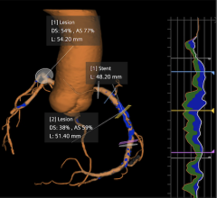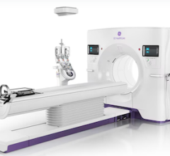
A 3-D reconstruction made from an image acquired by a Philips Healthcare CT system.
Computed tomography (CT) scanner manufacturers say they are tracking numerous trends in the cardiac imaging market that are influencing their development efforts. Among them are concerns over ionizing radiation dose, increasing use of CT to evaluate chest pain patients, development of CT perfusion imaging and the need to simplify cardiac imaging and workflow.
“What facilities are telling us is they want to be able to scan the largest number of patients at the lowest possible dose,” said Jamie Valliant, senior director, Philips Healthcare head of global product marketing for CT.
He said this has been made possible with a combination of software, hardware and simplification of operation. From the software side, he said vendors are extracting more image data than ever before, allowing low-dose acquisitions to be processed into diagnostic-quality images. “What we as vendors are asking is how we can extract as much information as we can from each photon we collect,” Valliant said.
“Cardiac CT has improved dramatically over the last few years with the technical advances that have been made,” said Joseph Cooper, director of Toshiba’s CT business unit. “Eventually I think CT will become the gold-standard, because it’s less expensive and noninvasive.”
Evaluating Chest Pain, Cutting Costs
CT is gaining ground as a fast, noninvasive chest pain screening tool for hospital emergency rooms. “From the patient’s perspective, all they have is an IV in the arm and a three-second scan,” Cooper said.
Vendors and cardiac CT experts contend coronary CT angiography (CCTA) will eventually replace conventional diagnostic catheterization angiography and nuclear myocardial perfusion imaging (MPI). CT screening can help cut healthcare costs incurred using the current standards of enzyme blood test, electrocardiogram (ECG), ECG stress test, perfusion imaging and then diagnostic catheterization, said Guillaume Grousset, Siemens product marketing manager for dual source CT. He said CT is ideal at chest pain centers to quickly screen low- and medium-risk patients to rule out a heart attack, because CT has a high negative predictive value.
“It will take a while, but there are a lot of financial studies showing the economic impact of using CT,” Grousset said. “The amount of clinical data showing this is the right thing to do is overwhelming.”
He said this will reduce patient volume in the cath lab, but in the current political climate of reducing healthcare costs, cardiac CT will likely become a very attractive option. Grousset suspects cardiac CT will be reimbursed accordingly by the Centers for Medicare and Medicaid Services (CMS) and private insurance companies to encourage its use.
In September, results from the Coronary Computed Tomographic Angiography for Systematic Triage of Acute Chest Pain Patients to Treatment (CT-STAT) trial were published in the Journal of the American College of Cardiology. Data suggest that employing early CCTA is faster, more accurate and less costly than employing rest-stress MPI in the evaluation of acute low-risk chest pain in the emergency department. Low-risk patients were randomized to treatment via CCTA or MPI and followed up over a period of six months. The two treatments were analyzed primarily over the time taken to diagnose and the cost of care to the emergency department.
The results found that CCTA patients were diagnosed 54 percent faster than MPI patients and the total costs of care were 38 percent lower with the CCTA group. Major adverse cardiac events were no different for each diagnostic strategy. The CCTA patients were also exposed to less radiation than the MPI patients (11.5 vs 12.8 mSv).
Since about 8 million U.S. patients require emergency department evaluation for acute chest pain annually, widespread CCTA use could have a major impact on costs, said DeAnn Haas, RT (R) (CT) (MR), GE Heathcare marketing manager of CT segment.
Cooper predicts lower dose, increased resolution and accuracy and the cost-effectiveness of CT over catheterization and MPI will help expand the appropriate use criteria for cardiac CT.
The Next Step: CT Myocardial Perfusion Imaging
A major advance on the horizon for CT is myocardial perfusion imaging. CT is excellent at imaging anatomy, but the question of whether an occlusion in a vessel merits treatment has been left to follow-up nuclear perfusion imaging or evaluation in a cath lab using catheter-based fractional flow reserve (FFR) measurements. If clinical trial data can prove cardiac CT is also accurate at imaging perfusion, it might eliminate the need for additional nuclear imaging scans or diagnostic catheterizations.
“We have gotten to the point where we can show the anatomy very well, but now CT may be able to provide FFR and perfusion information as well,” said Haas.
Haas said FFR-CT is being developed by HeartFlow, a California software company. The technology is being evaluated in the DeFACTO (Determination of Fractional Flow Reserve by Anatomic Computed Tomographic Angiography) trial. The prospective, multi-center trial, conducted at up to 20 U.S., Canadian, European and Asian centers, is designed to determine the diagnostic performance of HeartFlow’s CT-FLOW software by CCTA for noninvasive assessment of the hemodynamic significance of coronary lesions, as compared to direct measurement of FFR during cardiac catheterization as a reference standard.
Recent hardware advances, especially larger detector sizes, have also enabled CT perfusion imaging, since the whole heart and its blood flow can be frozen in one moment in time. For example, Cooper said Toshiba’s 320-slice scanner can capture the entire heart in less than one second and offers better temporal resolution without stitching or motion artifacts from lower-slice systems. He explained the speed and large imaging area of the 320-slice scanner allow imaging of perfusion through the organ and wash out to identify areas of ischemia. “It really is a game-changing technology,” Cooper said.
However, compelling clinical trial data is needed to prove CT perfusion imaging has clinical utility over the standard of nuclear MPI and/or invasive diagnostic cath lab angiography procedures. Cooper said the Combined Noninvasive Coronary Angiography and Myocardial Perfusion Imaging Using 320 Detector Computed Tomography (Core320) trial was developed to show how CT fares against both diagnostic catheter angiography and CT perfusion versus SPECT perfusion imaging.
“We hope to show cardiac CT is more effective and cost-effective than the traditional procedures,” Cooper said.
Reducing Dose
Patient radiation dose from X-ray imaging technologies such as CT has been at the forefront of radiology concerns since popular media has shed light on several cases of extreme overexposure of patients who received CT exams.
Part of the problem has been the size of CT slices, which could not cover the entire heart in one beat, requiring several images to be taken and stitched together to reconstruct a complete image of the heart. To do this, scanners used retrospective ECG gating, where the CT scanner was left on for several cardiac cycles and data from several segments were used to piece together a complete image. However, this required a larger dose, up to 30-35 mSv, Grousset said.
“That’s about 10 times the annual amount of background radiation the patient receives,” he explained. “You throw away a lot of your data, so you are giving radiation dose to the patient for images you will not use.”
Today, most CT systems use prospective ECG gating to trigger the X-ray source only when it will capture images that are needed. He said this can reduce dose tremendously, down to about 3-5 mSv. Newer systems can now perform cardiac scans in a quarter of a second, with dose down to 1 mSv or less.
“The focus on radiation dose has been good for the industry because it has made the vendors innovate with dose-lowering technology,” Cooper said. “There has been a dramatic reduction in radiation dose with prospective gating, and that is going to get better with iterative reconstruction software.”
Software improvements have been the biggest contributor to lowering CT dose. Over the past seven years, all vendors have developed their own versions of iterative reconstruction software, which filters the noise out of low-dose scans to improve image quality, said Mani Vembar, Philips Healthcare CT clinical scientist.
The Need for Speed
Cardiac imaging is all about speed (temporal resolution) to freeze the motion of a moving organ, Grousset said.
“Everything being done in cardiac CT is being done to get images of the coronary arteries, and those are fast-moving objects,” he explained. “Ideally, you want to freeze motion in one heart beat.”
One way to do this is by increasing temporal resolution, which is the time it takes for a CT scanner to create a slice. For example, if a whole rotation of the CT gantry takes one second to create a slice, this can be cut to half a second using data from half a rotation with reconstruction software.
“You need a fast scan and can use temporal resolution to help,” Grousset said. “Until recently there were no CT systems that could freeze motion.”
The average 64-slice CT scanner used for cardiac imaging cannot image the entire heart in one scan. Increasing the number of slices a scanner can acquire at one time can help freeze this motion. Vendors have introduced 256- and 320-slice scanners capable of imaging the entire heart in one scan in less than a second. Toshiba’s Aquilion One 320-slice CT scanner can acquire 16 cm in one scan, in one phase of the cardiac cycle, in less than a second. The average length of a heart is about 12 cm.
Faster exam speeds enable motion to be frozen, as long as a patient’s heart rate is 60 beats or fewer, Grousset said.
Dual-source CT scanners have also helped reduce dose and increase speed. Siemens says its dual-source CT system has an average cardiac exam dose of 0.84 mSv and an exam time of 75 milliseconds. The dual detectors also mean scans can be performed in a quarter rotation, instead of a half rotation.
Haas said more speed and slices are nice, but the cost of higher-slice systems may be a limiting factor for many facilities’ budgets. She said there also is a question of whether there are better patient outcomes to justify these costs as compared to 64-slice scanners.
Automating Cardiac Imaging
Post-processing software developments over the past few years enable perfusion imaging and automated removal of surrounding anatomy from data sets so only the heart or coronary arteries are shown to speed diagnosis. Software can also take cardiac data sets and automatically calculate ejection fractions and label anatomy and specific vessels.
“The job of the cardiologist is not to make 200 clicks to create an image; they are there to make a diagnosis of those images. This software helps them get to that diagnosis faster,” Grousset said.
Another software innovation will be computer-aided detection for heart disease. Siemens is developing auto-detection software to not only find plaque in coronary vessels, but also to evaluate the degree of stenosis.
Another trend in CT systems is simplifying these complicated systems so radiology techs can produce better quality images, said Vembar.
What the Future Holds
In addition to CT myocardial perfusion imaging, transcatheter aortic valve implantation (TAVI) planning and new post-processing software will be the next major advances in cardiac CT.
Grousset said CT image data sets are critical in helping optimize outcomes in TAVI. CT imaging is first used to screen patients who are suitable for the procedure or to see if they should be referred for surgical repair. The CT data is used to plan the access route to ensure the larger-sized catheters can navigate the vessels to the heart. Finally, he said CT data is used to size the valve that will be used and verify proper replacement post-procedure.
Comparison Chart
This story was an introduction to a comparison chart for CT systems marketed specificially for cardiac imaging. To find the chart, click on the "comparison charts" tab at the top of the page. Participants in the chart include:
- GE Healthcare, www.gehealthcare.com
- Hitachi, www.hitachimed.com
- Philips Healthcare, www.philips.com/healthcare
- Siemens Healthcare, www.usa.siemens.com/flash
- Toshiba, medical.toshiba.com


 February 02, 2026
