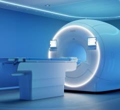
Siemens SOMATOM Definition Flash dual-source CT is among the new CT systems that can significantly cut dose and imaging times through speed. New CT reimbursements are expected to boost interest in cardiac CT in 2010.
With new CPT reimbursement codes for cardiac computed tomography (CT) starting in January 2010, there will likely be renewed interest in CT this coming year.
This and other imaging market insights were offered by Christian Renaudin, D.V.M., Ph.D., MBA, managing partner and CEO of The MarkeTech Group LLC. The company performs market research for medical-technology companies, including some of the largest imaging device manufacturers.
Computed Tomography
In cardiac CT Dr. Renaudin said there will be an increase in 128 and 256-slice scanners to help decrease procedure times and to increase resolution. However, he said so far there are no good clinical studies showing these high-slice systems are any better at diagnostics than a typical 64-slice system. He expects sales will do better when these studies begin to surface.
One set back in CT is in overall sales, which Dr. Renaudin said dropped by 20-30 percent in the United States over the past year due to the recession initially and now with the uncertainty of the pending healthcare reform.
He said the U.S. market is widely seen as the engine for purchases in the worldwide imaging market. Until the U.S. sales pickup he expects the CT market to remain flat worldwide.
The lack of cardiac CT reimbursement has been a major issue holding back the modality, Dr. Renaudin said. “Some modalities are not well valued by payers,” he said, including cardiac CT and MRI.
However, the American Medical Association announced in October it created four new Category 1 CPT codes for cardiac CT, which become effective Jan. 1, 2010. The new codes offer a universal insurance billing code for cardiac CT, which previously did not exist and opens the door to reimbursements in 2010.
This move will likely change the attitudes toward CT and open the likelihood of its expanded use in cardiology.
The increased use of CT imaging has raised concern about exposing patients to increased radiation. Dr. Renaudin said there is a trend to find ways to reduce radiation, including faster, higher-slice CT scanners.
Faster scanners will result in the need for new, faster contrast media injectors that can keep up with the pace. He said these new injectors will be designed in ways to help reduce the amount of contrast agent that is used and will better integrate with EMRs. This evolution will include more dual-chamber injectors, because single chamber injectors cannot keep up with the faster speeds and require bolus and saline flush protocols.
MRI vs. SPECT
“For years we have been predicting cardiac SPECT would die and cardiac MRI would take off,” Renaudin said. “I think the predicted death of SPECT is going to be delayed for another 10 years.”
He describes MRI as a one-stop shop for cardiac imaging because it can provide the same anatomical imaging as CT, the same information as echo, and the same perfusion imaging as SPECT nuclear scans. MRI also does not expose the patient to any radiation. However, he admits MRI is expensive and is not easily accessible. He said the economics for dedicated cardiac MRI are just not there, so for now SPECT imaging will likely remain the dominant perfusion imaging system. He said SPECT gamma cameras are also cheap compared to MRI systems and are available for in-office use, while MRI siting cost is too expensive to serve as a office-base machine.
The quality of SPECT imaging is also improving, he added. In June 2009 GE Healthcare released its Alcyone Technology, a nuclear cardiology SPECT platform combining cadmium zinc telluride (CZT) detectors, focused pin-hole collimation, 3D reconstruction and stationary data acquisition. GE said the technology is designed to improve workflow, dose management and overall image quality. GE claims that Discovery NM/CT 570c will cut scanning times from 20 minutes to three minutes. Other equivalent technologies already exist in the market (Digirad, Spectrum Dynamics) and it is still early to be able to compare the different products. Dr. Renaudin said the new technology could hurt echo and MRI because it is faster and has better throughput.
SPECT/CT and PET/CT
The manufacturers of the new combined SPECT/CT and PET/CT imaging systems say these systems offer better attenuation correction and hybrid images with better anatomical landmarks to increase diagnostic accuracy. However, many cardiologists say new software can correct attenuation without the large, extra cost of a built-in CT scanner, according to Renaudin. In addition, many cardiologists say they do not need CT’s anatomical landmarks because they already know what part of the heart they are looking at. CT guidance is more valued with oncology applications.
For these reasons Dr. Renaudin said SPECT/CT and PET/CT systems have been a hard sell for the cardiology market. He said they are better suited for imaging cancer, which is reflected in the fact that 95 percent of the imaging studies performed by these scanners are oncology exams.
Echocardiography
The biggest advancement in the past couple years for echocardiography has been the introduction of 3D and 4D imaging, primarily for evaluating heart structure, such as valve function. Dr. Renaudin said 3D echo is still a top-tier hospital modality because of its expense. But, he said the prices are falling with increased competition in the market.
He said ultrasound is not just about the machine, it’s also about the user and their skill in interpreting the images. He sees a trend where vendors are attempting to create analytical software to help interpret the images.
“There is a greater effort to bring in artificial intelligence and take the guesswork out of the analysis,” Renaudin said.
He said progress in this area is much further along in CT imaging, but is not there yet with echo. One of the things the analytical systems need to do before they will be able to match CT interpretive systems is take an image and convert it into standardized indexes of numbers and measurements for things like ejection fractions.
Angiography
Among the big advances in digital angiography X-ray systems is a move to create CT-like images in the cath lab. The major angiography system manufacturers have each created their own versions of CT-like imaging where the C-arm does a rotation around the patient on the table and creates a 3D image. The image can be used for overlays on angiography for better navigation, anatomical reference, and can help visualize stent positioning.
Dr. Renaudin points out one of the shortcomings of angiography is that it does not show the true lumen wall in vessels with stenosis. However, the CT-like images can help reveal the true anatomy. He said the biggest advantage of these new tools will likely be in better mapping vessels in the brain. This road-mapping solution already exists in the neuro cath lab.

 May 14, 2026