
The annual Radiological Society of North America (RSNA) scientific assembly and annual meeting is where imaging systems vendors showcase their latest technology. While aimed at the radiology market, imaging systems specific to cardiovascular specialties are often highlighted at RSNA first.
Latest CT Advances
Philips, GE and Siemens unveiled their next generation computed tomography (CT) systems, each offering significant advances over technology these vendors currently offer. A new generation of CT scanners is being readied to produce angiograms, captured at high speeds and paradoxically low radiation dose, that might allow more accurate planning and effective implantation of stents, as well as repair heart valves; for example, transcatheter aortic valve implantations. Such exams are possible now only with Toshiba’s path-finding Aquilion One, but soon competitors from Siemens and GE, shown as works in progress at this year’s RSNA meeting, will enter the market.
The GE Revolution CT can capture a human heart in a single beat free from motion artifact. At the show, the company framed the 510(k) pending system as an “all-in-one” scanner, offering coverage, and spatial and temporal resolution that can handle a comprehensive range of radiological applications, while serving as a platform for new applications.
GE said the Revolution platform is not just an upgraded CT system, as every component was entirely redesigned from the ground up using the latest technologies to increase speed, improve image quality and reduce dose. It offers 256 detector rows and features an 80 cm bore and a gantry rotation speed of 0.28 seconds. It can perform a cardiac CT angiography (CTA) scan in less than a second with less than 1 mSv dose. The Revolution CT features GE’s new ASiR-V dose reduction technology, which is a hybrid of GE’s ASiR iterative reconstruction software and its model base reconstruction software, Veo.
Siemens billed its Somatom Force as the next generation in dual-source CT. Successor to the Somatom Definition Flash, the company’s previous top-tier CT, Force features two generators that — like the generators on its predecessor — fires X-rays independently at variable energies. Force is so fast, however, that it can image the adult chest in a second, eliminating the need to breathhold, and freeze a human heart pumping at 90 beats per second by merging the two imaging chains to create a 50 cm (nearly 19.7-inch) field of view. Alternatively the two imaging chains, like those on the Flash, can be tuned to separate energies to differentiate among tissues or materials, as in visualizing iodinated contrast as it flows through the coronaries or accumulates in varying amounts in benign versus malignant masses, such as those in the kidney.
The Somatom Force has the potential to allow use of significantly less iodine contrast than in the past and can perform chest scans without the need for breath holds. The two X-ray generators on Siemens’ Somatom Force can be pumped at low voltage (as low as 70 kVp) to deliver low-dose scans. Dose can be reduced further using the company’s SAFIRE software.
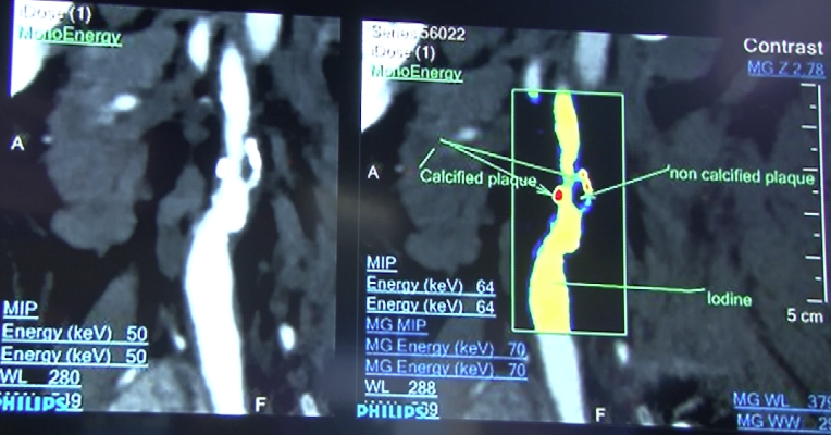
Newly unveiled at RSNA 2013 and 510(k)-pending, the IQon Spectral CT from Philips attempts such differentiation using a single imaging chain to capture X-rays at different wavelengths. In the same way that white light is made up of a spectrum of colors to that relate to chemical elements, the X-ray beam photons used in CT scanners also consist of a spectrum of X-ray energies. With the development of a new yttrium-based spectral detector that can discriminate between X-ray photons of multiple high and low energies simultaneously, IQon can deliver both anatomical information and the ability to characterize structures based on their chemical makeup within a single scan.
Using a type of spectral analysis, the system can separate out materials made up of specific atomic numbers off the periodic chart of elements. The system so far has been tested for iodine and calcium. This can be used to help differentiate between areas of high blood contrast uptake and calcified areas, which can be useful in diagnosing kidney stones and better delineating various types of atherosclerotic plaque in arteries. Elements can be assigned specific color codes to make them stand ut on scans, even if the surrounding tissue has similar Hounsfield unit numbers. The spectral imaging features are still pending 510(k) approval, but the vendor hopes the U.S. Food and Drug Administration (FDA) will review the technology in 2014.
Angiography Advances
At RSNA 2012, GE and Siemens introduced all-new premium angiography system platforms using the latest low-dose technology. Siemens introduced its new Artis Z and Artis Zen platforms that use new types of X-ray tubes and detectors for very low dose acquisitions. The Siemens’ systems also introduced new software tools to better track devices and guide coronary interventions. GE featured its Interventional Guidance System (IGS) platform, which departs from traditional fixed mounting of high-quality C-arms to the floor or ceiling, instead using a fully motorized, wireless rolling gantry that uses laser guidance to move about the room. This year, Siemens and GE introduced mid-range price versions of these new angiography platforms, the Siemens’ Artis One and GE’s IGS 740.
The Artis One is designed to fit within a 269-square-foot room, has an installation time of four days and uses 20 percent less energy consumption than previous generations of Siemens angiography systems.
Philips followed this year with the introduction of its new Allura Clarity angiography platform, which uses new software algorithms with faster computers to enhance lower dose images by correcting motion, reducing noise and reducing pixel shifting for better cine imaging. The enhancements help improve image resolution of smaller vessels and at a lower dose.
Toshiba highlighted three dose reduction technologies for its Infinix-i cardiovascular imaging systems. This included a real-time angiography X-ray patient dose monitoring system that highlights where on the body the dose is being concentrated. This shows physicians where the dose is being concentrated during long procedures so they are aware and can move the C-arm to minimize damage such as skin burns.
Toshiba also showed a novel approach to minimizing dose. The vendor’s Spotfluoro minimizes radiation exposure by surrounding a rectangular region of interest (ROI) with a “last-image hold,” an image captured some seconds earlier. Updating the image in the ROI exposes the patient to less dose, just as it keeps the interventionalist current on what is happening.
Xbox Has a Place in the Cath Lab/Hybrid OR
New Xbox gaming technology allows players to use hand movement gestures to zoom in and out and scroll through a menu. This technology is now being translated into practical use to view 3-D anatomical images in operating rooms (OR) and interventional suites. Two German start-up companies demonstrated this technology at RSNA 2013 — Christian Schaller, founder and CEO of the German tech start-up Metrilus, and professor Michael Teistler, Ph.D., creator of the Teistler Imager.
Using time-of-flight 3-D cameras similar to technology built into the Xbox Kinect motion sensor, Schaller was able to call up an image by pointing to it with a single finger, switch displayed images with the swipe of a hand, and zoom an image by motioning up and down.
Teistler used an off-the-shelf USB plug-and-play Xbox controller to quickly scroll through 3-D CT reconstructions of the heart, choose an image and then rotate, zoom and flip through slices of the image on any access to see the detailed coronary anatomy.
The interfaces are both prototypes and a long way from application. But the allure is clear for physicians being able to easily manipulate 3-D anatomical reconstructions on screen during a procedure.
Gesture navigation offers surgeons the promise of remote control over display technology without physical contact. The advantage is obvious to anyone who has tried to maneuver the tight spaces of an operating theater, much of which is draped in blue, signifying scrubbed and sterilized surfaces that better not be touched.
Imaging to Aid Vessel Access
Evena Medical showed a new hands-free technology for use in cath labs to visualize the blood vessels up to 10 mm under the skin for easier vascular access. The Evena Sparrow system consists of a small tablet-sized screen with a multi-spectral light source underneath that emits multiple wavelengths to clearly visualize the vessels as dark lines on the skin seen on the screen. A second-generation system, Evena Glasses, eliminates the need for the screen device and instead combines the light source, cameras and recording capability into a pair of glasses worn by the clinician.
The system can clearly visualize femoral and radial arteries, immediately enables verification of vessel patency and gives immediate visual detection of extravasation/infiltration. Images can be recorded and downloaded for use in electronic documentation of cases.
The vendor said Stanford University Hospital’s cath lab was an early adopter of this technology.
Nuclear Imaging
Siemens shone a spotlight on its Biograph mCT Flow PET/CT scanner for improvements in image quality and standard uptake values (SUV) thanks to a continuously moving patient table that ushers body areas steadily through the detector, producing a more reproducible and robust stream of data. The company’s Symbia Intevo SPECT/CT leveraged the CT data set to create more accurate SPECT images, as well as combining the two data sets to allow, for the first time, accurate quantitation on a SPECT system.
What mCT Flow could do at the premium end of molecular imaging, Symbia Intevo might do for the masses performing bread-and-butter nuclear scans. Early adopters will see the benefit in bone scans, as Siemens has honed the technology first for this application. Other applications are expected to follow.
Philips further extended the power of positron imaging with a solid-state replacement for the photomultiplier tubes that have registered photons in PET scanners since their development in the 1970s. Philips positioned the new detector as the core of the industry’s first truly digital PET/CT, the company’s new Vereos. The new system, already cleared by the FDA, is truly remarkable as a pathfinder technology and a watershed development for Philips, which was late to market with a PET/CT but now may have an edge, as the PET-installed base prepares to replace aging systems. The company claims its detector produces images with two times better image resolution than PMT-based scanners and SUV measurements that are twice as accurate.
GE showed a work in progress PET/MRI system to match the systems previously released by Philips and Siemens. The prototype combines a GE MR750 3.0T system into a single, compact gantry with a PET detector. GE hopes to submit the system for FDA 510(k) review in 2014.
Quiet MRI
Noise pollution, a problem in the magnetic resonance imaging (MRI) suite since the very beginning of this modality, has been addressed mostly through the use of squishable ear plugs, handed out by techs. Wedge those chunks the wrong way and the bizarre noises that characterize MRI are a patient’s constant — and very loud — companions. Last year GE unveiled Silent Scan, a partial solution to the problem for one MRI sequence, a solution the company expanded this year to four more sequences. While Silent Scan is anything but silent, it helps. So much, in fact, that Siemens followed suit with its QuietSuite, which — like Silent Scan — reduces the noise made by the body coil that surrounds the patient.
Point-of-Care Ultrasound
Konica Minolta launched the pocket-sized Sonimage P3 hand-held ultrasound device, which is in the same class and low price range of GE Healthcare’s Vscan, introduced in 2010. It brings advanced imaging capabilities to the patient or bedside in an imaging system small enough to fit in a lab coat pocket or can be worn like a stethoscope. It can help clinicians at the point of care visually assess a patient immediately for a rapid diagnosis.
The durable, palm-sized device weighs less than 14 ounces and offers B-mode, M-mode and Doppler in an easy-to-use operational format and presets. The Sonimage P3 can be used as a stand-alone ultrasound system or plugged into a Windows-based PC, laptop or tablet for the flexibility of a larger display. All Sonimage P3 devices come standard with Sonimage View software for online imaging and offline review and storage. The device also features a touch screen, choice of interchangeable transducers and a 4 GB data card that stores more than 10,000 images.
Samsung has made major strides in the past two years to rapidly build itself as a new but substantial player in the radiology market. The vendor showed its work in progress UGEO WS80A ultrasound system featuring “5-D” imaging that automatically extracts relevant anatomical views for a study from the volumetric data acquired by the tech. Its initial application is for OB/GYN exams, but may have application in cardiology, which currently requires experienced techs to extract specific views, which sometimes results in operator variability.
Technology Trends Into the Future
The annual RSNA meeting is a barometer for new trends in imaging and imaging related information technology.
A big trend in imaging has been the increase in quantification of anatomy. Quantification turns subjective observations into objective measures upon which physicians can base patient management decisions. Advanced cardiac quantification packages, many with automation features to greatly speed workflow, today can be found on nearly all cardiac ultrasound systems and in MRI, CT, echo and nuclear imaging software.
Vendors have met some of information technology challenges posed by Stage 2 Meaningful Use that call for the use of remote image viewing systems and improved patient engagement through technology. The number of vendors offering remote viewing systems has exploded over the past year. These systems enable easy sharing of medical images via e-mail, portals or direct connections to PACS systems for referring physicians, or image access via electronic medical records (EMRs).
Several vendors this year showed patient portals to allow them access to their electronic medical records, labs, billing information, scheduling, radiology images and exam or procedural reports. Web-based patient portals allow increased patient engagement to more actively include them in their healthcare management.
Another big trend at RSNA 2013 was the introduction of several software solutions to monitor and track patient radiation dose from X-ray sources, especially CT and angiography. There has been increasing interest in recording dose levels since several states have passed or are considering laws requiring hospitals to record X-ray doses. It also is widely expected that appropriate dose level guidelines will eventually be set by medical societies in the future.
For a video overview of the key trends and latest technologies at RSNA 2013, watch the video of the Editor’s Choice of the Most Innovative New Technologies.
Editor’s note: Greg Freiherr has reported on developments in radiology since 1983. He runs the consulting service, The Freiherr Group. Read more of his views on his blog at itnonline.com.


 June 17, 2026


