
The annual Radiological Society of North America (RSNA) scientific assembly and annual meeting is where imaging systems ...
Computed Tomography (CT)
Cardiac computed tomography CT systems use a series of X-ray images to create an image volume dataset that can be sliced or manipulated on any plane using advanced visualization software. This channel includes content on CT scanners, CT contrast agents, CT angiography (CTA and CCTA), CT perfusion, spectral CT (also called dual souce or dual energy CT), and interative image reconstruction software that can reduce dose and make lower-quality CT images diagnostic.

Feature | Dave Fornell
DAIC readers chose the following stories as the most popular content in 2013, based on website analytics. The list is ...
 January 03, 2014
December 31, 2013 — Massachusetts was on the same brink in 2006 that the entire nation is on today: the brink of ...
December 31, 2013Sponsored Content

Blog | Enterprise Imaging
As medical advancements continue to push the boundaries of what is possible in the field of structural heart ...
August 10, 2023
December 26, 2013 — A study shows that coronary artery calcium (CAC) screening, an assessment tool that is not currently ...
December 26, 2013Following public comment received in the fall of 2013, The Joint Commission has released new accreditation standards for ...
December 20, 2013
December 17, 2013 — The American College of Radiology (ACR) commends the House Committee on Ways and Means and the ...
December 17, 2013December 17, 2013 — A new study by researchers at the University of California, Los Angeles (UCLA) Fielding School of ...
December 17, 2013By Dave Fornell, DAIC editor
While the Radiological Society of North America (RSNA) focuses on new technology for ...
December 16, 2013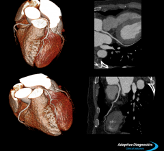
December 12, 2013 — Toshiba introduced its Adaptive Diagnostics computed tomography (CT) technology, which simplifies ...
December 12, 2013December 11, 2013 — The U.S. Centers for Medicare & Medicaid Services (CMS) finalized updates to payment policies and ...
December 11, 2013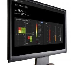
December 10, 2013 — GE Healthcare showcased several new offerings that enable low-dose imaging procedures and help ...
December 10, 2013
Hear why Siemens SOMATOM Definition Edge is the CT your emergency department (ED) has been dreaming about from the ...
December 10, 2013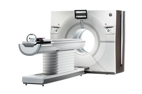
December 9, 2013 — GE Healthcare announced at the Radiological Society of North America Annual Meeting (RSNA 2013) in ...
December 09, 2013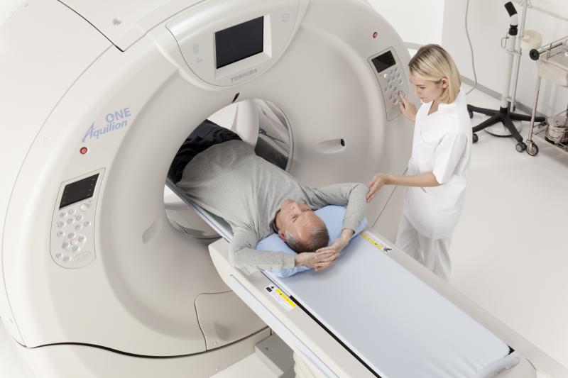
December 6, 2013 — Over the past decade, medical imaging has gone from being one of the fastest growing categories of ...
December 06, 2013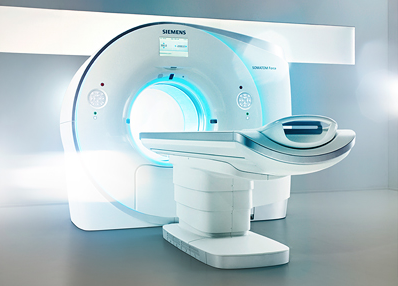
December 2, 2013 — Siemens Healthcare introduced Somatom Force, the company’s next-generation Dual Source computed ...
December 02, 2013