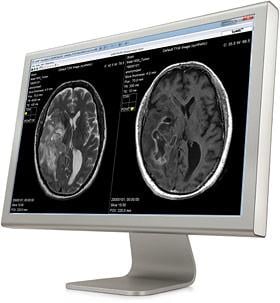
Image courtesy of SyntheticMR AB
June 2, 2015 — Cincinnati Children’s Hospital Medical Center (CCHMC) has evaluated synthetic magnetic resonance imaging (MRI) on pediatric cases to study and clinically validate its use on children. Results from the first part of the evaluation, which used SyMRI software, shows the synthetic images are diagnostically satisfactory in comparison with conventional sequences.
Long examination times are particularly challenging when examining children, who may even need to be given a general anesthetic during the exam. A faster workflow would therefore be especially valuable for pediatric applications. CCHMC sees an opportunity to improve the efficiency of pediatric MRI by being able to shorten the examination time with SyMRI. Synthetic imaging is being evaluated on pediatric cases at CCHMC in two steps. At first it has been ensured that the synthetic images are diagnostically sufficient in comparison with conventional sequences.
SyntheticMR AB develops and markets new MRI software solutions. It created the SyMRI Image software that provides fast MRI workflows, allowing high patient throughput, and SyMRI Neuro that enables automatic segmentation of brain tissue, providing objective decision support based on quantitative data. SyMRI is a CE-marked product. A customized version of SyMRI Image was introduced at the 2014 Radiological Society of North America (RSNA) annual meeting by SyntheticMR’s strategic partner GE Healthcare under the name MAGiC for GE’s line of Signa MRI systems.
“Because of the way the brain develops, a true strength of synthetic imaging that could be particularly useful in pediatric cases is the possibility of tuning image contrast afterwards and generating contrasts that usually are not acquired. Therefore the use of synthetic imaging, including interactive contrast changes during review, is now ongoing as the second step of evaluation. The ability to create multiple diagnostic sequences from a single acquisition may allow for substantial time savings and increased throughput”, said Blaise V. Jones, M.D., chief of neuroradiology, Cincinnati Children’s Hospital Medical Center.
While clinical validation of synthetic MRI relative to standard pulse sequences has been performed in adults, no such data has previously been available on children. The study at CCHMC evaluates synthetic MRI in children by comparing synthetic MR images with conventional MR images in children at both 1.5 and 3.0T in a blinded fashion to determine image quality and diagnostic efficacy relative to standard MRI.
“We see great potential for its use in clinical practice, and anticipate that synthetic imaging is going to help us discern subtle differences at grey matter to white matter interfaces, for example in epilepsy cases. Furthermore we expect that the quantitative T1, T2 and PD maps can assist us in monitoring the myelination process of white matter during the first years of brain development”, Jones continued.
SyMRI IMAGE from SyntheticMR optimizes the MRI workflow and shortens scan time using synthetic MRI. This is achieved by a single MR quantification scan of 5-7 minutes. Based on this single scan, conventional standard T1W, T2W and FLAIR images and any other combination of TE, TR and TI can be shown without rescanning the patient, allowing a substantial reduction in examination time.
For more information: www.symri.com

 May 14, 2026