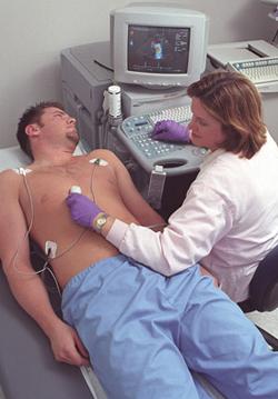
October 3, 2011 – Up to now, on-the-job hazards for cardiac sonographers were usually limited to overtaxed muscles or incorrect body positioning. And the profession, with help from equipment manufacturers, has made remarkable progress in identifying and lessening these problems. But now, based on discussions initiated on the American Society of Echocardiography’s (ASE) social media website, [email protected] and results of an ASE survey, there appear to be growing concerns about radiation exposure. As October is Medical Ultrasound Awareness Month, the ASE believes it is appropriate to address this emerging concern for cardiovascular sonographers. Are sonographers getting more radiation on the job than is acceptable for their health?
Initial results of a member poll, which includes responses from 90 sonographers, confirms that 63 percent feel that radiation safety is of concern in their workplaces, yet only 13 percent of these respondents say that a formal policy was in place to address radiation safety for cardiovascular sonographers. “I feel that this is an important topic of concern. I have been doing echo in an outpatient setting for 17 years, and when I’ve raised this topic with my managers and physicians, I felt that they have not wanted to address the subject. I have implemented a ‘loose’ policy that somewhat addresses safety by doing the echo before the patient is injected with a radioisotope, but I’m in close quarters with these patients every day. I recently did a literature search on this topic, but found few relevant articles. I absolutely feel this needs closer review,” said one survey respondent. Additionally, the preliminary survey information indicates that not all institutions treat exposure risks the same way.
As echocardiography is a safe, radiation-free diagnostic test, how are sonographers getting exposed? In 2006, Americans were exposed to more than seven times as much ionizing radiation from medical procedures as was the case in the early 1980s, according to a 2009 report on population exposure by the National Council on Radiation Protection and Measurements (NCRP). Medical exposure constituted nearly half of the total radiation exposure of the U.S. population from all sources in 2006, and was primarily a result of the growth in the use of medical imaging procedures, including computed tomography (CT) and nuclear medicine.
As more cardiovascular laboratories move toward a multi-modality model where nuclear and ultrasound imaging are done in the same facility, and as more sonographers work in these multi-modality environments or integrated outpatient centers, there may be a need to protect them from an unacceptable level of exposure. In ASE’s poll, 98 percent of the respondents say that both echocardiograms and nuclear medicine studies are performed in their facilities, and 21 percent of them regularly spend time in the catheterization lab.
How much is too much? The Health Physics Society (HPS), which is dedicated to making sure that health physicists have the information necessary to manage the beneficial use of ionizing radiation while protecting workers and the public from potential hazards, suggests that close contact with radiated patients can quickly lead to an unacceptable level of exposure. Significant radiation exposure can be acquired from being close to patients who have both nuclear stress tests and cardiovascular ultrasound on the same day, and from spending time in catheterization labs and hybrid rooms.
The Health Physics Society has advised putting personal radiation monitors (body badges) on sonographers to find out how much dose they actually receive per patient. ASE sonographer Sue Maisey, RDCS, FASE, director of non-invasive cardiology/arrhythmia at St. Luke’s Episcopal Hospital/Texas Heart Institute in Houston, reports that all sonographers at her institution wear radiation badges, which are sent out for monitoring each month. Their exposure rates are posted and reviewed by the hospital radiation safety officer. In the ASE’s survey, there was a wide variety of procedures for handling patients who receive both nuclear and ultrasound tests on the same day, ranging from facilities that schedule all ultrasounds before patients are radiated and monitor sonographers for radiation exposure, to facilities where changing the schedule to avoid exposure is considered inconvenient to patients or staff and strongly resisted, and those where no radiation monitoring is done.
Sonographer Marti McCulloch, RDCS, MBA, FASE, director of Methodist DeBakey Heart & Vascular Center in Houston, notes that at her institution, the growing volume of patients who receive both echocardiography and nuclear stress tests on the same day has led to the scheduling of echocardiograms prior to nuclear tests when possible. She stated,“Radioisotopes vary according to test and gamma camera, so it is important for both the patient and the sonographer staff to be aware of what types of tests have been recently performed on the patient.” She recommends that sonographers talk with their hospital administrators to put in place policies that facilitate having patient tests be routed correctly, e.g., ultrasound tests being performed on patients first, before the nuclear department exams.
ASE believes there is a need for a greater awareness of the causes of radiation exposure and adherence to prevention techniques for the working sonographer. ASE is continuing to collect information on this issue and gathering best practices for addressing concerns; resource information available to date has been posted on ASE’s website. ASE will partner with other sonographer and ultrasound organizations to make any information as widely available as possible. In the member poll, a number of respondents favored creation of a uniform radiation policy; some suggested that standards set by ASE and/or the Intersocietal Commission on the Accreditation of Echocardiography Laboratories (ICAEL) would have more credibility in the workplace than those created by labs independently.
For more information: www.aesecho.org

 June 17, 2026


