December 1, 2015 — Athletes who engage in the extreme sport of free diving, descending hundreds of feet below the surface of the ocean while holding their breath, undergo significant cardiovascular changes, according to a new study presented today at the annual meeting of the Radiological Society of North America (RSNA). These changes can pose potential dangers, particularly to inexperienced or cardiac untrained divers.
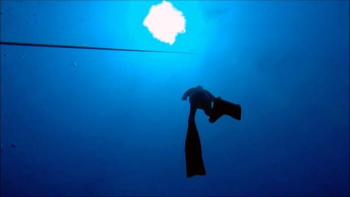
Apnea is the temporary suspension of breathing. The ancient practice of free or apnea diving has experienced immense growth worldwide over the past decade, due to coverage in the media and increased competition and training opportunities for elite and recreational divers. The sport can be dangerous, because divers must hold their breath for prolonged periods while undergoing massive water pressure and physiological changes.
Recreational divers are at greatest risk because of lack of conditioning, but even elite divers have suffered lasting or fatal effects resulting from free diving. Most recently, champion diver Natalia Molchanova was reported missing and presumed dead off the coast of Spain during a dive in August 2015.
Researchers at the University of Bonn in Bonn, Germany, used MRI to study the simulated effects of free diving on the cardiovascular systems of 17 elite free divers from Germany and Austria (age range 23 to 58). To study the effects of a lack of oxygen on heart function and blood flow, respectively, the divers underwent cardiac MRI and MRI of the carotid arteries before, during and after a maximum breath hold.
"We wanted to look at the changes that occur in the heart during apnea in real time," explained study author Jonas Dörner, M.D., who is now a radiology resident at the University Hospital of Cologne.
The average apnea was 299 seconds (just under five minutes) and 279 seconds or about four and a half minutes for the first and second MRI exams, respectively. The maximum breath hold (or apnea) during the exams was eight minutes and three seconds.
"These athletes train to be able to hold their breath for long periods," Dörner said. "When they get into the water, they are able to hold their breath even longer due to the diving reflex."
When submerged underwater without access to oxygen, the body responds with what is called "diving reflex," which includes a decreased heart rate, a constriction of blood vessels in the extremities, and a shift in blood flow from the extremities to the brain. These changes also occur – to a lesser degree – during prolonged breath holding without being submerged. As oxygenated blood is diverted from the rest of the body to the brain, blood pressure increases.
The MRI exams allowed the researchers to observe the cardiovascular changes involved in the diving reflex in real time. During apnea, the amount of blood flowing to the brain through the carotid arteries increased and then leveled off.
"At the beginning of the apnea period, the heart pumped more strongly than when the heart was at rest," Dörner said. "Over time, the heart dilated and began to struggle."
By the end of the apnea period, Dörner said the divers' heart function began to fail.
"At that point, not enough blood is being pumped to the brain," he said. "The heart is unable to pump against the high resistance of the blood vessels."
Although the changes in the divers' systolic heart function during apnea are similar to those in patients with systolic heart failure, Dörner said that the condition was transient in the divers.
"The divers' heart function recovered within minutes of breathing again," said Claas Nähle, M.D., head of this cardiac magnetic resonance research group. "It appears that elite divers develop compensatory mechanisms that help them adapt to the cardiovascular changes that occur during apnea." However, for individuals with less training, free diving may be problematic.
"As a recreational activity, free diving could be harmful for someone who has heart or other medical conditions and is not well trained for the activity," said one of the study's leaders, Lars Eichhorn, M.D., from the Department of Anesthesiology and Intensive Care Medicine at the University Hospital of Bonn.
Eichhorn added that deaths among highly trained divers are mostly seen in the discipline called deep diving, a special type of apnea diving that combines the risk of prolonged apnea with changes of ambient pressure.
Other co-authors on the study are Jean-Marc Lunkenheimer, Julian A. Luetkens, M.D., Juergen Gieseke, D.Sc., Rainer Meyer, Ph.D., Andreas Hoeft, M.D., and Hans H. Schild, M.D.
For more information: RadiologyInfo.org

 January 28, 2026