New cutpoints for identifying adolescents and young adults at risk of an enlarged heart and premature cardiac damage. Image courtesy of Andrew Agbaje
March 9, 2023 — An enlarged heart, often described as left ventricular hypertrophy, is a sign of early cardiac damage which has been linked to long-term cardiovascular diseases and death in adults. In children and adolescents, left ventricular hypertrophy is often recognised as a consequence of elevated blood pressure and metabolic disorders.
Echocardiography can be used to assess left ventricular hypertrophy. The most accurate echocardiography measurement is left ventricular mass indexed for muscle mass. This requires a dual energy-Xray absorptiomery scan of muscle mass which is often not available due to cost. Other indices for left ventricular mass such as height and body surface area are useful although less accurate.
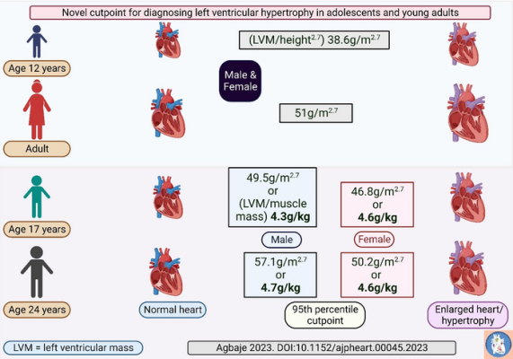
The most widely used cutpoint for diagnosing left ventricular hypertrophy in children and adolescents was established over 30 years ago and the average age of the children participating in that study was about 12 years. The established cutpoint identified left ventricular mass indexed for height greater than 38.6g/m2.7 as left ventricular hypertrophy in a pediatric population. The 38.6g/m2.7 is the 95th percentile value, meaning that 95% of children remain below this value, and it corresponded to 3.4g/kg of left ventricular mass indexed for muscle mass.
Among adults, the established cut point for diagnosing left ventricular hypertrophy is 51g/m2.7. Until now, there have been no large enough studies to define an accurate cutpoint for diagnosing left ventricular hypertrophy during late adolescence and young adulthood. The current pediatric cutpoint is significantly low for mid- and late-adolescents and the adults’ cut point is quite high, both leading to the misclassification of adolescents with normal cardiac mass as abnormal.
The current study was conducted among 868 adolescents who were 17 years old and followed up for 7 years until young adulthood at age 24 years. All 868 adolescents had dual-energy Xray absorptiometry body composition measurements and echocardiography measurements at baseline and follow-up.
The results revealed that during growth from age 17 years to 24 years, muscle mass and left ventricular mass increased significantly in both males and females, although more in males. However, when left ventricular mass was indexed for total body muscle mass, it removed the difference in the cardiac mass between the males and females by young adulthood.
The researchers also observed that estimating left ventricular mass by height2.7 or 3 had the best agreement with left ventricular mass divided by muscle mass. The left ventricular mass indexed for muscle mass 95th percentile cutpoint for males and females was 4.3g/kg and 4.6g/kg respectively at age 17 years. At 24 years of age, the 95th percentile cutpoints were 4.7g/kg and 4.6g/kg for males and females, respectively.
Correspondingly, the 95th percentile cutpoint for left ventricular mass indexed for height reflecting left ventricular hypertrophy in males and females was 49.5g/m2.7 and 46.8g/m2.7 respectively at age 17 years. At 24 years of age, the 95th percentile cutpoints were 57.1g/m2.7 and 50.2g/m2.7 for males and females, respectively.
“These new cutpoints for the young population are higher than the pediatric cutpoint and lower than the adult cutpoint, except among 24 year old males. Thus, an extra 10 – 20g/m2.7 left ventricular mass indexed for height in comparison to the currently used pediatric cutpoint may be normal in adolescents and young adults, depending on sex. Therefore, pediatricians, cardiologists, physiologists, researchers, and caregivers are encouraged to familiarize themselves with the new cutpoints in order to accurately stratify adolescents and young adults at risk of an enlarged heart and premature cardiac damage,” says Andrew Agbaje, a physician and clinical epidemiologist at the University of Eastern Finland.
Dr Agbaje’s research group (urFIT-child) is supported by research grants from Jenny and Antti Wihuri Foundation, the Finnish Cultural Foundation Central Fund, the Finnish Cultural Foundation North Savo Regional Fund, the Orion Research Foundation sr, the Aarne Koskelo Foundation, the Antti and Tyyne Soininen Foundation, the Paulo Foundation, the Yrjö Jahnsson Foundation, the Paavo Nurmi Foundation, the Finnish Foundation for Cardiovascular Research and the Foundation for Pediatric Research.
For more information: https://www.uef.fi/en

 June 12, 2026


