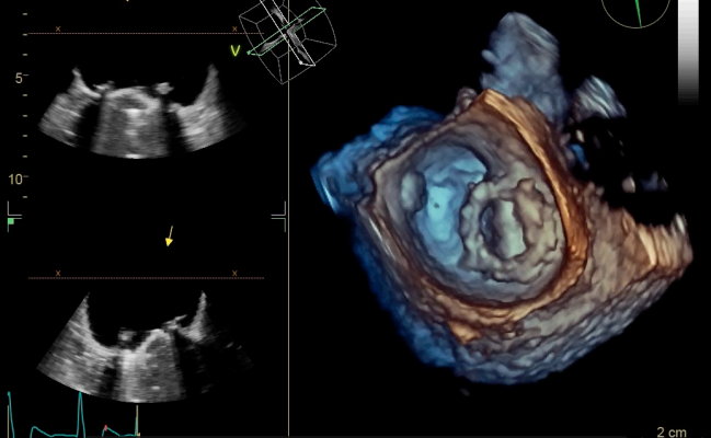
This case study is from the Cardiac Imaging Department, Hospital Clinic, Barcelona, Spain. A woman in her early 60s came to the emergency room with left hemiparesis and her medical history suggested several reasons for concern. She was a sufferer of rheumatic valve disease since childhood and had a history of valve surgeries. They used 3-D echo to clearly identify a small thrombus covering part of the prosthetic ring, which had not been completely clear in the 2-D echo.
File
MRI scanner_from DeBakey heart.jpg464.53 KB

 April 30, 2026