
With improved clinical capabilities for cardiac mapping and navigation during EP procedures, EnSite System Version 6 is designed to provide 3-D cardiac models at the time of an EP procedure.
Physicians create the 3-D model in small sections that join together automatically, resulting in a cardiac model that shows the openings, ridges and curves in the heart. The models are then used by physicians to collect information about the heart's electrical activity and assist them in diagnosing and treating many arrhythmias, including atrial fibrillation.
Frequently used features are accessible from the main screen and images can be displayed at full-screen size for greater visibility.
Technology | June 28, 2006
3-D Cardiac Models Provided at Time of EP Procedure
Additional Content
Sept. 2, 2025 — Stereotaxis has announced the successful completion of the world’s first procedures using MAGiC Sweep, a ...
 September 05, 2025

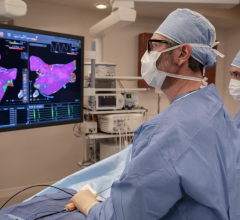
May 15, 2023 — Biosense Webster, Inc., a global leader in cardiac arrhythmia treatment and part of Johnson & Johnson ...
May 15, 2023 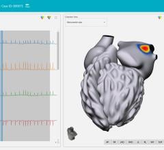
April 29, 2022 – Vektor Medical has announced positive results from its “Vektor vMap Clinical Validation Study” ...
April 29, 2022 
March 3, 2022 – CIRCA Scientific, Inc. has announced that its S-CATH M Esophageal Temperature Probe is now cleared for ...
March 03, 2022 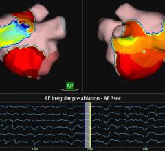
An example of the Acutus Medical AcQMap High Resolution Imaging and Mapping System to guide electrophysiology (EP) ...
October 14, 2021