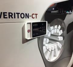
April 14, 2010 – The Mount Sinai Medical Center in New York has become the first facility in the United States to commercially use a new cardiac nuclear imaging system that drastically reduces imaging time and radiation exposure.
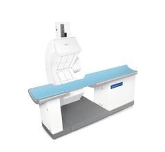
The new Discovery NM 530c single photon emission computed tomography (SPECT) scanner uses a cadmium zinc telluride (CZT)-based, high-speed, high-resolution camera.
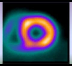
“The device provides detailed pictures of the heart that enable Mount Sinai physicians to quickly and more accurately assess the location, extent, and severity of heart disease,” said Milena J. Henzlova, M.D., professor of medicine (cardiology), Mount Sinai School of Medicine. “The new CZT-based nuclear cameras have already made a big difference in evaluating our patients for coronary artery disease. In addition to significantly reducing patient radiation exposure and increasing the number of patients we can examine each day, this technology provides a cost-effective way for us to diagnose heart disease quickly and with confidence so that patients receive treatment sooner.”
With conventional nuclear cardiac imaging, patients must hold their arms above their head for two cardiac scans that take between 15-20 minutes each. With the Discovery NM 530c, the scanning time is reduced to three to five minutes for each scan. This reduction can be more comfortable for a patient, and it can possibly reduce imaging artifacts caused by patient movement. A shorter, more comfortable scan has the opportunity to improve image quality, allowing Mount Sinai clinicians to be more confident in their diagnosis.
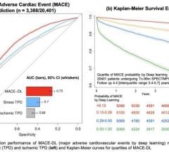
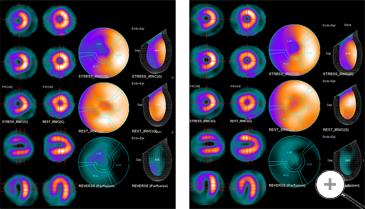
At the recent American College of Cardiology annual scientific session in Atlanta (ACC.10), Dr. Henzlova and colleagues presented data showing that the new CZT camera technology also improves cardiac imaging while reducing radiation exposure. Patients in their study were divided into three groups based on the type of test they received to evaluate their heart health: a cardiac stress test with a low isotope dose; a cardiac stress test with a high isotope dose; and the traditional two part test where the patient is given a low dose of radiation while resting and a high dose during a stress test. Researchers compared the image quality using all three different isotope doses using the new camera.
Results showed that the proportion of patients with excellent or good image quality was similar in all three groups (91-98 percent). However, compared to the traditional testing in the rest-stress group, radiation was decreased on average by 70 percent in the low dose stress-only group and by 30 percent in the high dose stress-only group.
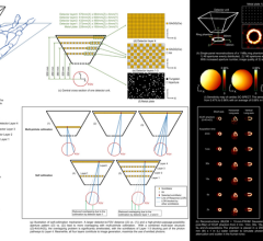
The scanner technology consists of four key elements:
• CZT detectors improve image resolution.
• Focused pin-hole collimation is used for improved detection efficiency, resulting in greater image clarity and speed.
• Stationary data acquisition is used to acquire all views simultaneously during a fully stationary SPECT acquisition. This huge decrease in moving parts virtually eliminates the risk of motion artifacts and significantly shortens scan times.
• 3-D reconstruction is used to generate accurate and easily interpretable images of the myocardial region.
The Mount Sinai Medical Center is the first laboratory in the United States to receive accreditation for the use of the camera from the Intersocietal Accreditation Commission. Mount Sinai currently has two Discovery NM 530c units in use.
For more information: www.mountsinai.org
 June 05, 2023