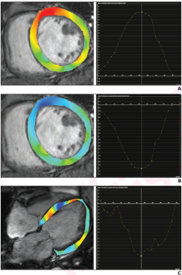
80-year-old patient with nonischemic dilated cardiomyopathy. Ejection fraction was 25.2%. Patient underwent cardiac MRI. A. Radial strain overlay on short-axis cine image and corresponding graph show global radial strain measurement, yielding value of 10.8%. B. Circumferential strain overlay on short-axis cine image and graph show global circumscribed strain measurement, yielding value of -8.5%. C. Longitudinal strain overlay on 4-chamber cine image and graph show global longitudinal strain measurement, yielding value of -10.7%. Global longitudinal strain measurement indicates lesser myocardial strain. Patient did not experience major adverse cardiovascular event after 3 years of follow-up.
November 9, 2022 — According to an accepted manuscript published in ARRS’ American Journal of Roentgenology (AJR), left ventricular global longitudinal strain—derived from feature tracking on cardiac MRI—is a significant independent predictor of adverse outcomes in patients with dilated cardiomyopathy.
“CardioThis study strengthens the body of evidence supporting the clinical implementation of feature tracking when performing cardiac MRI in patients with dilated cardiomyopathy,” wrote corresponding author Dr. Ming-Yen Ng from the department of diagnostic radiology at the University of Hong Kong.
In this AJR accepted manuscript, 471 patients (365 men, 106 women; median age, 61 years) with ischemic (n=233) or nonischemic (n=238) dilated cardiomyopathy and left ventricular ejection fraction <50% underwent cardiac MRI at four centers from January 2011 to December 2019. Manual contouring helped to determine cardiac MRI parameters; additionally, software-based feature tracking was used to calculate six myocardial strain parameters: left ventricular (LV) and right ventricular (RV) global radial, global circumferential, and global longitudinal strain. Late gadolinium enhancement (LGE) was also evaluated.
Ultimately, in patients with dilated cardiomyopathy who underwent cardiac MRI with feature tracking, LV global longitudinal strain was a significant predictor of a composite outcome of all-cause mortality and/or heart-failurehospitalization (hazard ratio=1.13, p=.006), independent of age, corrected LV and RV end-diastolic volume, LV and RV ejection fraction, and presence of late gadolinium enhancement.
Noting that the literature has published conflicting results regarding the utility of cardiac MRI-derived feature tracking parameters, “this multicenter study supports the role of LV global longitudinal strain as a prognostic marker of adverse outcomes in patients with dilated cardiomyopathy,” the authors of this AJR accepted manuscript reiterated.
For more information: www.arrs.org

 May 21, 2026


