September 22, 2009 – Toshiba America Medical Systems Inc. yesterday introduced Low-Contrast Imaging (LCI) for Toshiba’s Infinix-i systems with mid (12” x 12”) and large (12” x 16”) flat-panel detectors (FPD).
Toshiba will demonstrate its low-contrast imaging capabilities at this year’s Transcatheter Cardiovascular Therapeutics (TCT) annual meeting in San Francisco, Sept. 21-25, at its booth 705.
The company said it enables better visualization of soft tissue to improve the imaging and treatment of patients by offering CT-like images of soft tissue, including brain tissue, cerebral ventricles and hepatic visualization, without requiring additional exams.
Beneficial for both physicians and patients, low-contrast imaging allows better visualization of soft tissue in the angiography suite and enables more accurate diagnosis of a variety of diseases using a single system. Additionally, this technology may be used to confirm appropriate endpoints during interventional procedures, such as aortic stent-grafting. Capturing CT-like images without the use of an additional test helps improve accuracy and workflow in the angio suite. It also leads to lowering overall health care costs by using a single system to treat numerous diseases and handle multiple procedures.
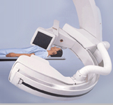
The Infinix-i line features a five-axis C-arm that enables head-to-toe and fingertip-to-fingertip coverage and allows greater clinician access to the patient for diagnostic and interventional procedures. The freely moving components, ergonomically friendly design and five-axis positioner enable physicians to obtain optimal angles for cardiac diagnosis and interventional procedures without repositioning the patient. Infinix’s unique, flexible design improves workflow and collaboration between cardiologists, interventional cardiologists, anesthesiologists and clinical staff during exams, especially in hybrid OR settings.
For more information: www.medical.toshiba.com

 June 12, 2026


