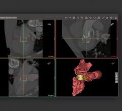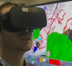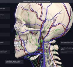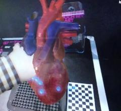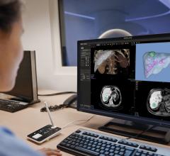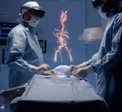
An example of 3-D computer aided design (CAD) software from Materialise being used at Henry Ford Hospital to evaluate left ventricular outflow tract (LVOT) obstruction from a Sapian TAVR valve virtually implanted in the mitral valve position. Henry Ford uses both 3-D printing and CAD to plan and guide complex structural heart procedures.
With increasing complexity of interventional structural heart disease and congenital heart disease interventions, 3-D printing of the anatomy is being used for pre-planning and practicing procedures, device sizing and as a visual reference for the soft tissue during procedures. 3-D printing has become more popular within the medical space as it has been shown to help prevent, fix and foresee procedural errors.
Many lather hospitals are integrated 3-D printing services to better personalized patient care. These centers have 3-D printing labs to create accurate anatomical models from a patient's computed tomography (CT) scans.
However, printed models of the heart show one momement frozen in time from a dynamic organ that changes shape and twists throughout the cardiac cycle. For this reason, some centers are now also using computed aided design (CAD) software that can virtually model the anatomy during the entire cardiac cycle. This enables more accurate valve sizing and to model what happens when a device is placed in a moving heart.
Uses of 3D Printing in Cardiology
In the setting of paravalvular leaks around failed surgical devices, many times patients are not candidates for a second or third redo operation due to progressive underlying frailty. The leaks are often plugged using off-label transcatheter occluder devices. These highly complex procedures once took five to seven hours in duration, but 3-D printed models of patient-specific anatomy now offers a better understanding of the leaks and can help preplan procedures with device sizing and how these occluder devices will interact with adjacent anatomy. Use of 3-D models can decrease procedural time by up to a half.
A 2018 study by the University of Minnesota in Minneapolis examined the effectiveness of 3-D printing and computer modeling to predict paravalvular leak (PVL) in patients undergoing transcatheter aortic valve replacement (TAVR). A common risk of TAVR is an ill-fitting valve which can lead to PVL. The study used 3-D printing technology to help confirm and detect the location of the leak. CT scans allowed researchers to see a 360-degree view of the location of the calcium build up while the 3D models allowed researchers to further evaluate the ill-fitting valves. The 3D aortic root models were then virtually implanted with the valve to determine if the size was correct and revealing where there were leaks around the areas of heavy calcium. The models were ethen compared to in-vivo implanted TAVR echocardiograms.
Every leak seen on the 3D models were confirmed on the CT digital scans. The 3D models allowed researchers to use prototypes to personalize valve placement, size and location to stop leaks and lower calcium build up.
“We are very encouraged to see such positive outcomes for the feasibility of 3D printing in patients with heart valve disease. These patients are at a high risk of developing a leak after TAVR, and anything we can do to identify and prevent these leaks from happening is certainly helpful,” said lead author Sergey Gurevich, M.D., assistant professor of medicine, Cardiovascular Division at the University of Minnesota in Minneapolis, Minn. “Like any other new technology, as 3D printing evolves, we hope to see an increase in accessibility and opportunity for the use of this technology to help improve patient care.”
For transcatheter left atrial appendage (LAA) occlusion procedures, 3-D printing can be used to understand the best location of a transeptal puncture to enable the delivery catheter to enter the LAA, explained Dee Dee Wang, M.D., director, structural heart imaging at Henry Ford Hospital, Detroit. She said these models also can help determine which catheters are best suited to that particular patient's anatomy. This preplanning can save procedure time and the need to exchange multiple devices.
"It allows us to be able to feel a patient's heart that we otherwise could not, because we are not surgeons, and that tactile sensation and concept of special resolution in being able to turn the heart outside the body, really gives us a better idea of the procedure before we go into it," Wang explained.
3-D Printed Hearts Help Guide Complex Procedures
Another use case for in-house medical 3-D printing is to create surgical models to help cardiac surgeons plan complex cases. This is especially true with multidisciplinary cases in interventional radiology, cancer surgery and pediatric cardiac surgery. These types of models are used at Florida-based Nemours Children’s Health System as a pre-planning blueprint and roadmap for surgeons and proceduralists. The hospital said these help increase confidence, reduce procedure times and minimizing unexpected findings while in the operating room.
“The simulation aspect of 3-D modeling is a game changer. To be able to look at a model of a tumor from all angles, without the restrictions of a 2-D image on a computer screen, is completely changing how we are planning complex surgery,” said Craig Johnson, DO, chair of medical imaging and enterprise director of interventional radiology.
Using modified models from volumetric 2-D CT and magnetic resonance imaging (MRI), physicians can run simulations of the surgery and more accurately determine the tools they will need, with the multidisciplinary team involved, cutting down on waste. For example, certain models enable surgeons to drill holes in them to measure and select the appropriate medical equipment to use during the procedure.
“Three-dimensional modeling prepares us by helping us know exactly what we’re going to do. We do not have to plan on the spot if we come across something unexpected. Instead we’ve had imaging from radiology as well and the model,” said Tamarah J. Westmoreland, M.D., Ph.D., a pediatric surgeon at Nemours Children’s Health System. “The surgery is almost like a musical concert. It is rehearsed, planned and then executed without complication.”
Nemours also found 3-D models are invaluable in explaining procedures to patients and their families. An echo, MRI or CTR can be difficult for a parent to conceptualize, so the Nemours team uses a true-to-size 3-D model of that patient. In many cases, the patients have the high-fidelity models next to them in their room as care teams explain their treatment plan.
“When we use a model to explain to a parent or a child a procedure, it’s clear, this approach is different,” Johnson explained. “They are able to visualize what we are going to do and it sets them at ease.”
Jessica Lewis is the mother of a Nemours patient and experienced this firsthand. Her 13-year-old son, Malachi, had a rare congenital coronary artery anomaly and needed cardiac surgery at Nemours.
“I was able to turn the model of my son’s artery around and look at it from all sides,” said Lewis. “The more educated you are about the procedure, the more empowered you feel because you completely understand what is going on with your child.”
Uses of 3D Computer Aided Design in Structural Heart Planning
One of the most import applications for CAD currently is to determine if there will be left ventricle outflow tract obstruction when placing a transcatheter metal valve. This is especially true with devices like the Edward Sapian transcatheter aortic valve replacement (TAVR) device used in the mitral valve position. The Sapian is frequently used in very sick patients with dysfunctional mitral valves who cannot undergo surgery, and currently there is no FDA cleared transcatheter mitral valve available.
She said experience with implanting Sapian in mitral valves, recording LVOT gradients and looking at post-procedure CTs allowed Henry Ford to developing computer modeling using CAD software from 3-D printing vendor Materialise to better predict which patients will have poor outcomes, or require more advanced procedures to better fit the valve using alcohol septal ablation or use of the LAMPOON procedure to cut the mitral leaflet to prevent LVOT obstruction.
"When we made a 3-D printed model it made perfect sense to us, but after doing so many prints we realized we can visualize the valve without having to actually physically print it," Wang said from Henry Ford Hospital's experience. "We do both computer modeling and printing so we can have better communication between the surgeon, implanter and the patient prior to the procedure. This enables everyone to speak the same language, because a surgeon may explain something differently than an interventionists who does not actually cut into the patient."
Henry Ford has now used 3-D printing and CAD in well over 1,000 patients.
She said CAD and 4-D CT imaging also has had a major impact on Henry Ford's LAA occlusion outcomes. "We have shown from our structural heart experience that using 3-d CT, 4-D CT and 3-D printing that we could actually have zero complications compared to the clinical trial average, which was 16 percent," Wang said. In addition to improving outcomes, she said they were able to reduce the number of devices used and the procedural time. "From a hospital administrator standpoint means a faster, more efficient lab. They can do more procedures, and for an implanter, they have the confidence they need to do the procedure."
Large Imaging Companies Partner With 3-D Vendors
As more hospitals use 3-D printing and CAD technologies, many medical imaging vendors have partnered with established 3-D vendors to offer their software integrated into their advanced visualization, PACS or enterprise imaging platforms.
An example of this include Philips integrating software from both 3D Systems and Stratasys, two global leaders in the 3-D printing industry, into its IntelliSpace Portal. The embedded 3-D modeling application can create and export 3-D models intuitively into the clinical workflow. The suite of clinician-focused rendering and editing tools helps assure the model reflects the true patient anatomy.
3D Systems’ precision healthcare capabilities include virtual reality simulators, 3-D-printed anatomical models, virtual surgical planning, patient-specific surgical guides, instrumentation and implants. Their solutions help to speed care and treatment and reduce costs.
Stratasys is a supplier of applied additive technology solutions, from 3-D-printed surgical planning models and medical device prototyping to advanced education and training. Stratasys' PolyJet-based full-color, multi-material 3-D printing solutions drive high-quality realism. Interfacing with Philips, customers can now rapidly design, order, and produce 3-D-printed anatomical structures on-demand from Stratasys Direct Manufacturing.
Some larger advanced visualization vendors offer partnerships with 3-D printing companies where STL (standard triangle language) files for printing can be created from medical imaging and then sent to an outside company for custom printing orders. This can save costs by outsourcing 3-D printing rather than deleting an in-hospital printing lab.
Other vendors have developed their own software or partnered with hospitals with advanced 3-D printing programs to integrate the technology into their own platforms. GE Healthcare is one of the vendors that has such software, allowing files for printing to be created from CT datasets using the AW Advanced Visualization software.
See and example of the GE technology for a hip fracture repair
First FDA Clearance for Diagnostic 3-D Anatomical Models
In August 2017, the U.S. Food and Drug Administration (FDA) announced that software intended to create output files used for printing 3-D patient-specific anatomical models used for diagnostic purposes is a Class II medical device and requires regulatory clearance.
The FDA has approved 3-D applications for medical use. The first FDA clearance came in March 2018 for Materialise NV became the first company in the world for 3-D printing anatomical models for diagnostic use. Materialise Mimics inPrint software is used for pre-operative planning and the fabrication of physical models for diagnostic purposes, including patient management, treatment and surgeon-to-surgeon communication.
Future of 3-D Printing May be Implantable Biological Tissues
Bioengineers at several academic centers have been refining 3-D printing technologies to print replacement biological tissues to implant in the human body. The eventual goal is in the future is to print replacement organs.
In the realm of cardiovascular medicine, a couple of these 3-D printing technologies to expect on the horizon soon will be bioprinted heart valves, of blood vessels for use in bypass procedures and tissue patches that might be used to repair areas of infarct.
This science fiction-like technology is rapidly developing. The biggest obstacle has been being able to print very detailed, complex structures, such as tissue vasculature. In 2019, bioengineers collaborating from Rice University, University of Washington, Duke University and Rowan University cleared a major hurdle on the path to 3-D printing replacement organs with a breakthrough technique that enabled exquisitely entangled vascular networks. The technique can mimic the body's natural passageways for blood, air, lymph and other vital fluids.
Layers are printed from a liquid pre-hydrogel solution that becomes a solid when exposed to blue light. A digital light processing projector shines light from below, displaying sequential 2-D slices of the structure at high resolution, with pixel sizes ranging from 10-50 microns. With each layer solidified in turn, an overhead arm raises the growing 3-D gel just enough to expose liquid to the next image from the projector.
"One of the biggest road blocks to generating functional tissue replacements has been our inability to print the complex vasculature that can supply nutrients to densely populated tissues," said bioengineer Jordan Miller, assistant professor of bioengineering at Rice University's Brown School of Engineering. "Further, our organs actually contain independent vascular networks — like the airways and blood vessels of the lung or the bile ducts and blood vessels in the liver. These interpenetrating networks are physically and biochemically entangled, and the architecture itself is intimately related to tissue function. Ours is the first bioprinting technology that addresses the challenge of multivascularization in a direct and comprehensive way."
Medical Grade 3D Printing Services and Printers Comparison Chart
This article served as an introduction for a comparison chart DAIC created for services and printers systems. Access the 3-D Printing Services and Printers Chart . This will require a login, but it is free and only takes a minute to fill out the form. It also will grant access to all 62 product comparison charts on the website.
Related 3-D Printing in Cardiology Content Content:
To Access the Online 3-D Printing and Printing Services Comparison Chart
VIDEO: Applications in Cardiology for 3-D Printing and Computer Aided Design
3-D Printed Heart Models Displayed at SCCT
Researchers 3-D Print Lifelike Heart Valve Models
Interventional Imagers: The Conductors of the Heart Team Orchestra
3D Systems Announces On Demand Anatomical Modeling Service
3-D Printed Models to Guide TAVR Improve Outcomes
Nemours Children's Health System Uses 3-D Printing to Deliver Personalized Care
Developments in Transcatheter Mitral Valve Replacement
1,000th 3-D Print Patient Treated at Henry Ford Health System
New Technique Allows More Complicated 3-D Bioprinting
Find more 3-D printing news and video

 May 12, 2020

